 Movie
Movie Controller

[English] 日本語
 Yorodumi
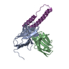
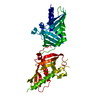
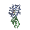
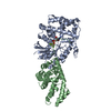
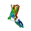
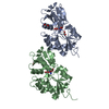
- PDB-6cf2: Crystal structure of HIV-1 Rev (residues 1-93)-RNA aptamer complex -
+ Open data

- Basic information
| Entry | Database: PDB / ID: 6cf2 | ||||||
|---|---|---|---|---|---|---|---|
| Title | Crystal structure of HIV-1 Rev (residues 1-93)-RNA aptamer complex | ||||||
 Components Components |
| ||||||
 Keywords Keywords | RNA BINDING PROTEIN/RNA / HIV-1 / Rev / RNA aptamer / RNA BINDING PROTEIN-RNA complex | ||||||
| Function / homology |  Function and homology information Function and homology informationhost cell nucleolus / viral process / mRNA transport / protein export from nucleus / host cell cytoplasm / DNA-binding transcription factor activity / RNA binding Similarity search - Function | ||||||
| Biological species |    Human immunodeficiency virus 1 Human immunodeficiency virus 1synthetic construct (others) | ||||||
| Method |  X-RAY DIFFRACTION / SYNCHROTRON / MOLECULAR REPLACEMENT / Resolution: 3 Å X-RAY DIFFRACTION / SYNCHROTRON / MOLECULAR REPLACEMENT / Resolution: 3 Å | ||||||
 Authors Authors | Eren, E. / Dearborn, A.D. / Wingfield, P.T. | ||||||
| Funding support |  United States, 1items United States, 1items
| ||||||
 Citation Citation | Journal: Structure / Year: 2018 Title: Structure of an RNA Aptamer that Can Inhibit HIV-1 by Blocking Rev-Cognate RNA (RRE) Binding and Rev-Rev Association. Authors: Dearborn, A.D. / Eren, E. / Watts, N.R. / Palmer, I.W. / Kaufman, J.D. / Steven, A.C. / Wingfield, P.T. | ||||||
| History |
|
- Structure visualization
| Structure viewer | Molecule: MolmilJmol/JSmol |
|---|
- Downloads & links
-Download
| PDBx/mmCIF format | 6cf2.cif.gz | 90.1 KB | Display | PDBx/mmCIF format |
|---|---|---|---|---|
| PDB format | pdb6cf2.ent.gz | 65.2 KB | Display | PDB format |
| PDBx/mmJSON format | 6cf2.json.gz | Tree view | PDBx/mmJSON format | |
| Others |  Other downloads Other downloads |
-Validation report
| Arichive directory | https://data.pdbj.org/pub/pdb/validation_reports/cf/6cf2ftp://data.pdbj.org/pub/pdb/validation_reports/cf/6cf2 | HTTPS FTP |
|---|
-Related structure data


-Links
 PDBj
PDBj


- Assembly
| Deposited unit | 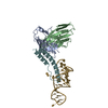
| ||||||||
|---|---|---|---|---|---|---|---|---|---|
| 1 |
| ||||||||
| Unit cell |
|
-Components
| #1: Antibody | Mass: 13408.822 Da / Num. of mol.: 1 / Fragment: Fab single-chain variable fragment Source method: isolated from a genetically manipulated source Source: (gene. exp.)  |
|---|---|
| #2: Antibody | Mass: 11656.958 Da / Num. of mol.: 1 / Fragment: Fab single-chain variable fragment Source method: isolated from a genetically manipulated source Source: (gene. exp.) |
| #3: Protein | Mass: 10746.137 Da / Num. of mol.: 1 / Fragment: UNP residues 1-93 Source method: isolated from a genetically manipulated source Source: (gene. exp.) Human immunodeficiency virus 1 / Gene: rev / Production host: |
| #4: RNA chain | Mass: 11275.749 Da / Num. of mol.: 1 / Source method: obtained synthetically / Source: (synth.) synthetic construct (others) |
| Has protein modification | Y |
-Experimental details
-Experiment
| Experiment | Method: X-RAY DIFFRACTION / Number of used crystals: 1 |
|---|
- Sample preparation
| Crystal | Density Matthews: 2.75 Å3/Da / Density % sol: 59.71 % |
|---|---|
| Crystal grow | Temperature: 291.15 K / Method: vapor diffusion, hanging drop / pH: 8 Details: 2% v/v 1,4-dioxane, 0.1 M Tris, pH 8.0, 15% w/v PEG3350 |
-Data collection
| Diffraction | Mean temperature: 100 K |
|---|---|
| Diffraction source | Source: SYNCHROTRON / Site: APS / Beamline: 22-ID / Wavelength: 1 Å |
| Detector | Type: MARMOSAIC 300 mm CCD / Detector: CCD / Date: Dec 16, 2016 |
| Radiation | Monochromator: double crystal Si(111) / Protocol: SINGLE WAVELENGTH / Monochromatic (M) / Laue (L): M / Scattering type: x-ray |
| Radiation wavelength | Wavelength: 1 Å / Relative weight: 1 |
| Reflection | Resolution: 3→38 Å / Num. obs: 10201 / % possible obs: 99.2 % / Redundancy: 2 % / CC1/2: 0.975 / Rmerge(I) obs: 0.088 / Rpim(I) all: 0.088 / Rrim(I) all: 0.124 / Net I/σ(I): 7.1 |
| Reflection shell | Resolution: 3→3.11 Å / Redundancy: 2 % / Rmerge(I) obs: 0.634 / Mean I/σ(I) obs: 1.7 / CC1/2: 0.424 / Rpim(I) all: 0.634 / Rrim(I) all: 0.897 / % possible all: 99.2 |
- Processing
| Software |
| ||||||||||||||||
|---|---|---|---|---|---|---|---|---|---|---|---|---|---|---|---|---|---|
| Refinement | Method to determine structure: MOLECULAR REPLACEMENT Starting model: PDB entries 5DHV & 1ULL Resolution: 3→38 Å / Cross valid method: FREE R-VALUE
| ||||||||||||||||
| Refinement step | Cycle: LAST / Resolution: 3→38 Å
| ||||||||||||||||
| LS refinement shell | Resolution: 3→3.158 Å
|
