 Movie
Movie Controller

[English] 日本語
 Yorodumi
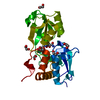
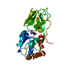
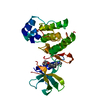
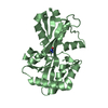
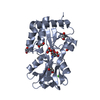
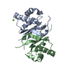
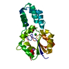
- PDB-5lom: Crystal structure of the PBP SocA from Agrobacterium tumefaciens ... -
+ Open data

- Basic information
| Entry | Database: PDB / ID: 5lom | ||||||
|---|---|---|---|---|---|---|---|
| Title | Crystal structure of the PBP SocA from Agrobacterium tumefaciens C58 in complex with DFG at 1.5 A resolution | ||||||
 Components Components | Deoxyfructosyl-amino Acid Transporter Periplasmic Binding Protein | ||||||
 Keywords Keywords | TRANSPORT PROTEIN / Periplasmic Binding Protein / Amadori compound / opine / DFG | ||||||
| Function / homology |  Function and homology information Function and homology informationligand-gated monoatomic ion channel activity / periplasmic space / membrane Similarity search - Function | ||||||
| Biological species |  Agrobacterium fabrum str. C58 (bacteria) Agrobacterium fabrum str. C58 (bacteria) | ||||||
| Method |  X-RAY DIFFRACTION / SYNCHROTRON / MOLECULAR REPLACEMENT / Resolution: 1.5 Å X-RAY DIFFRACTION / SYNCHROTRON / MOLECULAR REPLACEMENT / Resolution: 1.5 Å | ||||||
 Authors Authors | Marty, L. / Vigouroux, A. / Morera, S. | ||||||
 Citation Citation | Journal: J.Biol.Chem. / Year: 2016 Title: Structural Basis for High Specificity of Amadori Compound and Mannopine Opine Binding in Bacterial Pathogens. Authors: Marty, L. / Vigouroux, A. / Aumont-Nicaise, M. / Dessaux, Y. / Faure, D. / Morera, S. | ||||||
| History |
|
- Structure visualization
| Structure viewer | Molecule: MolmilJmol/JSmol |
|---|
- Downloads & links
-Download
| PDBx/mmCIF format | 5lom.cif.gz | 206.2 KB | Display | PDBx/mmCIF format |
|---|---|---|---|---|
| PDB format | pdb5lom.ent.gz | 164.7 KB | Display | PDB format |
| PDBx/mmJSON format | 5lom.json.gz | Tree view | PDBx/mmJSON format | |
| Others |  Other downloads Other downloads |
-Validation report
| Arichive directory | https://data.pdbj.org/pub/pdb/validation_reports/lo/5lomftp://data.pdbj.org/pub/pdb/validation_reports/lo/5lom | HTTPS FTP |
|---|
-Related structure data
| Related structure data |  5l9gC 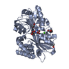 5l9iC 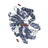 5l9lC  5l9oC 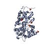 5l9pC  5l9sC  6hqhC  5l9m C: citing same article ( S: Starting model for refinement |
|---|---|
| Similar structure data |


-Links
 PDBj
PDBj
- Assembly
| Deposited unit | 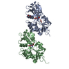
| ||||||||
|---|---|---|---|---|---|---|---|---|---|
| 1 | 
| ||||||||
| 2 | 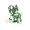
| ||||||||
| Unit cell |
|
-Components
| #1: Protein | Mass: 29010.648 Da / Num. of mol.: 2 / Fragment: UNP residues 36-278 Source method: isolated from a genetically manipulated source Source: (gene. exp.) Agrobacterium fabrum str. C58 (bacteria)Gene: socA, Atu5006 / Production host: #2: Chemical | 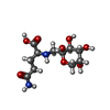  Mass: 308.285 Da / Num. of mol.: 2 / Source method: obtained synthetically / Formula: C11H20N2O8 Mass: 308.285 Da / Num. of mol.: 2 / Source method: obtained synthetically / Formula: C11H20N2O8#3: Chemical |   Mass: 62.068 Da / Num. of mol.: 3 / Source method: obtained synthetically / Formula: C2H6O2 Mass: 62.068 Da / Num. of mol.: 3 / Source method: obtained synthetically / Formula: C2H6O2#4: Water | ChemComp-HOH / |  Mass: 18.015 Da / Num. of mol.: 509 / Source method: isolated from a natural source / Formula: H2O Mass: 18.015 Da / Num. of mol.: 509 / Source method: isolated from a natural source / Formula: H2O |
|---|
-Experimental details
-Experiment
| Experiment | Method: X-RAY DIFFRACTION / Number of used crystals: 1 |
|---|
- Sample preparation
| Crystal | Density Matthews: 1.76 Å3/Da / Density % sol: 30.24 % |
|---|---|
| Crystal grow | Temperature: 292 K / Method: vapor diffusion, hanging drop / pH: 8.5 / Details: 30% PEG 4K, 100mM Tris |
-Data collection
| Diffraction | Mean temperature: 100 K |
|---|---|
| Diffraction source | Source: SYNCHROTRON / Site: SOLEIL  / Beamline: PROXIMA 2 / Wavelength: 0.9798 Å / Beamline: PROXIMA 2 / Wavelength: 0.9798 Å |
| Detector | Type: DECTRIS EIGER X 16M / Detector: PIXEL / Date: Jun 29, 2016 |
| Radiation | Protocol: SINGLE WAVELENGTH / Monochromatic (M) / Laue (L): M / Scattering type: x-ray |
| Radiation wavelength | Wavelength: 0.9798 Å / Relative weight: 1 |
| Reflection | Resolution: 1.5→50 Å / Num. obs: 64331 / % possible obs: 99.2 % / Redundancy: 6.71 % / Biso Wilson estimate: 18.27 Å2 / CC1/2: 0.997 / Rsym value: 0.01 / Net I/σ(I): 11.5 |
| Reflection shell | Resolution: 1.5→1.59 Å / Redundancy: 6 % / Rmerge(I) obs: 0.706 / Mean I/σ(I) obs: 2.1 / CC1/2: 0.856 / % possible all: 97.2 |
- Processing
| Software |
| ||||||||||||||||||||||||||||||||||||||||||||||||||||||||||||||||||||||||||||||||||||||||||||||||||||||||||||||||||
|---|---|---|---|---|---|---|---|---|---|---|---|---|---|---|---|---|---|---|---|---|---|---|---|---|---|---|---|---|---|---|---|---|---|---|---|---|---|---|---|---|---|---|---|---|---|---|---|---|---|---|---|---|---|---|---|---|---|---|---|---|---|---|---|---|---|---|---|---|---|---|---|---|---|---|---|---|---|---|---|---|---|---|---|---|---|---|---|---|---|---|---|---|---|---|---|---|---|---|---|---|---|---|---|---|---|---|---|---|---|---|---|---|---|---|---|
| Refinement | Method to determine structure: MOLECULAR REPLACEMENT Starting model: 5L9M 5l9m Resolution: 1.5→41.63 Å / Cor.coef. Fo:Fc: 0.953 / Cor.coef. Fo:Fc free: 0.939 / Rfactor Rfree error: 0 / SU R Cruickshank DPI: 0.087 / Cross valid method: THROUGHOUT / σ(F): 0 / SU R Blow DPI: 0.091 / SU Rfree Blow DPI: 0.089 / SU Rfree Cruickshank DPI: 0.087
| ||||||||||||||||||||||||||||||||||||||||||||||||||||||||||||||||||||||||||||||||||||||||||||||||||||||||||||||||||
| Displacement parameters | Biso mean: 19.96 Å2
| ||||||||||||||||||||||||||||||||||||||||||||||||||||||||||||||||||||||||||||||||||||||||||||||||||||||||||||||||||
| Refine analyze | Luzzati coordinate error obs: 0.21 Å | ||||||||||||||||||||||||||||||||||||||||||||||||||||||||||||||||||||||||||||||||||||||||||||||||||||||||||||||||||
| Refinement step | Cycle: LAST / Resolution: 1.5→41.63 Å
| ||||||||||||||||||||||||||||||||||||||||||||||||||||||||||||||||||||||||||||||||||||||||||||||||||||||||||||||||||
| Refine LS restraints |
| ||||||||||||||||||||||||||||||||||||||||||||||||||||||||||||||||||||||||||||||||||||||||||||||||||||||||||||||||||
| LS refinement shell | Resolution: 1.5→1.54 Å / Rfactor Rfree error: 0 / Total num. of bins used: 20
| ||||||||||||||||||||||||||||||||||||||||||||||||||||||||||||||||||||||||||||||||||||||||||||||||||||||||||||||||||
| Refinement TLS params. | Method: refined / Refine-ID: X-RAY DIFFRACTION
| ||||||||||||||||||||||||||||||||||||||||||||||||||||||||||||||||||||||||||||||||||||||||||||||||||||||||||||||||||
| Refinement TLS group |
|
