 Movie
Movie Controller

[English] 日本語
 Yorodumi
- PDB-5l9i: Crystal structure of the periplasmic binding protein MotA in comp... -
+ Open data

- Basic information
| Entry | Database: PDB / ID: 5l9i | ||||||
|---|---|---|---|---|---|---|---|
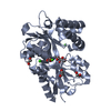
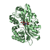
| Title | Crystal structure of the periplasmic binding protein MotA in complex with DFG from A. tumefaciens B6 | ||||||
 Components Components | periplasmic binding protein | ||||||
 Keywords Keywords | TRANSPORT PROTEIN / Periplasmic binding protein | ||||||
| Function / homology | Periplasmic binding protein-like II / D-Maltodextrin-Binding Protein; domain 2 / 3-Layer(aba) Sandwich / Alpha Beta / DI(HYDROXYETHYL)ETHER / Deoxyfructosylglutamine / :  Function and homology information Function and homology information | ||||||
| Biological species |  Agrobacterium tumefaciens str. B6 (bacteria) Agrobacterium tumefaciens str. B6 (bacteria) | ||||||
| Method |  X-RAY DIFFRACTION / SYNCHROTRON / MOLECULAR REPLACEMENT / Resolution: 1.9 Å X-RAY DIFFRACTION / SYNCHROTRON / MOLECULAR REPLACEMENT / Resolution: 1.9 Å | ||||||
 Authors Authors | Marty, L. / Morera, S. | ||||||
| Funding support |  France, 1items France, 1items
| ||||||
 Citation Citation | Journal: J.Biol.Chem. / Year: 2016 Title: Structural Basis for High Specificity of Amadori Compound and Mannopine Opine Binding in Bacterial Pathogens. Authors: Marty, L. / Vigouroux, A. / Aumont-Nicaise, M. / Dessaux, Y. / Faure, D. / Morera, S. | ||||||
| History |
|
- Structure visualization
| Structure viewer | Molecule: MolmilJmol/JSmol |
|---|
- Downloads & links
-Download
| PDBx/mmCIF format | 5l9i.cif.gz | 274.5 KB | Display | PDBx/mmCIF format |
|---|---|---|---|---|
| PDB format | pdb5l9i.ent.gz | 221.1 KB | Display | PDB format |
| PDBx/mmJSON format | 5l9i.json.gz | Tree view | PDBx/mmJSON format | |
| Others |  Other downloads Other downloads |
-Validation report
| Arichive directory | https://data.pdbj.org/pub/pdb/validation_reports/l9/5l9iftp://data.pdbj.org/pub/pdb/validation_reports/l9/5l9i | HTTPS FTP |
|---|
-Related structure data
| Related structure data |  5l9gSC 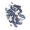 5l9lC  5l9oC 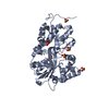 5l9pC  5l9sC  5lomC  6hqhC S: Starting model for refinement C: citing same article ( |
|---|---|
| Similar structure data |
-Links
 PDBj
PDBj- Assembly
| Deposited unit | 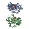
| ||||||||
|---|---|---|---|---|---|---|---|---|---|
| 1 | 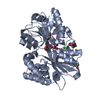
| ||||||||
| 2 | 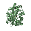
| ||||||||
| Unit cell |
|
-Components
-Protein , 1 types, 2 molecules AB
| #1: Protein | Mass: 38455.945 Da / Num. of mol.: 2 Source method: isolated from a genetically manipulated source Source: (gene. exp.) Agrobacterium tumefaciens str. B6 (bacteria)Gene: ASB65_13110 / Plasmid: pET28a / Production host: |
|---|
-Non-polymers , 5 types, 302 molecules 


| #2: Chemical |  Mass: 308.285 Da / Num. of mol.: 2 / Source method: obtained synthetically / Formula: C11H20N2O8 Mass: 308.285 Da / Num. of mol.: 2 / Source method: obtained synthetically / Formula: C11H20N2O8#3: Chemical |  Mass: 106.120 Da / Num. of mol.: 3 / Source method: obtained synthetically / Formula: C4H10O3 Mass: 106.120 Da / Num. of mol.: 3 / Source method: obtained synthetically / Formula: C4H10O3#4: Chemical | ChemComp-EDO /  Mass: 62.068 Da / Num. of mol.: 6 / Source method: obtained synthetically / Formula: C2H6O2 Mass: 62.068 Da / Num. of mol.: 6 / Source method: obtained synthetically / Formula: C2H6O2#5: Chemical |  Mass: 40.078 Da / Num. of mol.: 3 / Source method: obtained synthetically / Formula: Ca Mass: 40.078 Da / Num. of mol.: 3 / Source method: obtained synthetically / Formula: Ca#6: Water | ChemComp-HOH / | Mass: 18.015 Da / Num. of mol.: 288 / Source method: isolated from a natural source / Formula: H2O |
|---|
-Experimental details
-Experiment
| Experiment | Method: X-RAY DIFFRACTION / Number of used crystals: 1 |
|---|
- Sample preparation
| Crystal | Density Matthews: 2.22 Å3/Da / Density % sol: 44.62 % |
|---|---|
| Crystal grow | Temperature: 298 K / Method: vapor diffusion, hanging drop / pH: 6.5 / Details: 24% PEG 4K, 0.1 M MES, 0.2 M CaCl2 |
-Data collection
| Diffraction | Mean temperature: 100 K |
|---|---|
| Diffraction source | Source: SYNCHROTRON / Site: SOLEIL / Beamline: PROXIMA 1 / Wavelength: 0.97 Å |
| Detector | Type: DECTRIS PILATUS3 6M / Detector: PIXEL / Date: Feb 6, 2014 |
| Radiation | Protocol: SINGLE WAVELENGTH / Monochromatic (M) / Laue (L): M / Scattering type: x-ray |
| Radiation wavelength | Wavelength: 0.97 Å / Relative weight: 1 |
| Reflection | Resolution: 1.9→50 Å / Num. obs: 52621 / % possible obs: 99.7 % / Redundancy: 4.48 % / Biso Wilson estimate: 44.47 Å2 / Rsym value: 0.064 / Net I/σ(I): 11.9 |
| Reflection shell | Resolution: 1.9→2.01 Å / Rsym value: 0.888 |
- Processing
| Software |
| ||||||||||||||||||||||||||||||||||||||||||||||||||||||||||||||||||||||||||||||||||||||||||||||||||||||||||||||||||
|---|---|---|---|---|---|---|---|---|---|---|---|---|---|---|---|---|---|---|---|---|---|---|---|---|---|---|---|---|---|---|---|---|---|---|---|---|---|---|---|---|---|---|---|---|---|---|---|---|---|---|---|---|---|---|---|---|---|---|---|---|---|---|---|---|---|---|---|---|---|---|---|---|---|---|---|---|---|---|---|---|---|---|---|---|---|---|---|---|---|---|---|---|---|---|---|---|---|---|---|---|---|---|---|---|---|---|---|---|---|---|---|---|---|---|---|
| Refinement | Method to determine structure: MOLECULAR REPLACEMENT Starting model: 5L9G Resolution: 1.9→38 Å / Cor.coef. Fo:Fc: 0.96 / Cor.coef. Fo:Fc free: 0.949 / Rfactor Rfree error: 0 / SU R Cruickshank DPI: 0.14 / Cross valid method: THROUGHOUT / σ(F): 0 / SU R Blow DPI: 0.138 / SU Rfree Blow DPI: 0.123 / SU Rfree Cruickshank DPI: 0.125
| ||||||||||||||||||||||||||||||||||||||||||||||||||||||||||||||||||||||||||||||||||||||||||||||||||||||||||||||||||
| Displacement parameters | Biso mean: 52.47 Å2
| ||||||||||||||||||||||||||||||||||||||||||||||||||||||||||||||||||||||||||||||||||||||||||||||||||||||||||||||||||
| Refine analyze | Luzzati coordinate error obs: 0.24 Å | ||||||||||||||||||||||||||||||||||||||||||||||||||||||||||||||||||||||||||||||||||||||||||||||||||||||||||||||||||
| Refinement step | Cycle: 1 / Resolution: 1.9→38 Å
| ||||||||||||||||||||||||||||||||||||||||||||||||||||||||||||||||||||||||||||||||||||||||||||||||||||||||||||||||||
| Refine LS restraints |
| ||||||||||||||||||||||||||||||||||||||||||||||||||||||||||||||||||||||||||||||||||||||||||||||||||||||||||||||||||
| LS refinement shell | Resolution: 1.9→1.95 Å / Rfactor Rfree error: 0 / Total num. of bins used: 20
| ||||||||||||||||||||||||||||||||||||||||||||||||||||||||||||||||||||||||||||||||||||||||||||||||||||||||||||||||||
| Refinement TLS params. | Method: refined / Refine-ID: X-RAY DIFFRACTION
| ||||||||||||||||||||||||||||||||||||||||||||||||||||||||||||||||||||||||||||||||||||||||||||||||||||||||||||||||||
| Refinement TLS group |
|
