 Movie
Movie Controller


+ Open data

- Basic information
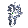
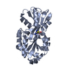
| Entry | Database: PDB / ID: 1wdn | ||||||
|---|---|---|---|---|---|---|---|
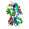
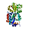
| Title | GLUTAMINE-BINDING PROTEIN | ||||||
 Components Components | GLUTAMINE BINDING PROTEIN | ||||||
 Keywords Keywords | COMPLEX (BINDING PROTEIN/PEPTIDE) / BINDING PROTEIN / GLNBP / CLOSED FORM / COMPLEX / COMPLEX (BINDING PROTEIN-PEPTIDE) / COMPLEX (BINDING PROTEIN-PEPTIDE) complex | ||||||
| Function / homology |  Function and homology information Function and homology informationL-glutamine binding / L-glutamine import across plasma membrane / L-glutamine transport / amino acid binding / amino acid transport / ligand-gated monoatomic ion channel activity / ATP-binding cassette (ABC) transporter complex, substrate-binding subunit-containing / outer membrane-bounded periplasmic space / periplasmic space / membrane Similarity search - Function | ||||||
| Biological species |  | ||||||
| Method |  X-RAY DIFFRACTION / Resolution: 1.94 Å X-RAY DIFFRACTION / Resolution: 1.94 Å | ||||||
 Authors Authors | Sun, Y.-J. / Rose, J. / Wang, B.-C. / Hsiao, C.-D. | ||||||
 Citation Citation | Journal: J.Mol.Biol. / Year: 1998 Title: The structure of glutamine-binding protein complexed with glutamine at 1.94 A resolution: comparisons with other amino acid binding proteins. Authors: Sun, Y.J. / Rose, J. / Wang, B.C. / Hsiao, C.D. #1: Journal: J.Mol.Biol. / Year: 1996Title: The Crystal Structure of Glutamine-Binding Protein from Escherichia Coli Authors: Hsiao, C.D. / Sun, Y.J. / Rose, J. / Wang, B.C. #2: Journal: J.Mol.Biol. / Year: 1994Title: Crystals of Glutamine-Binding Protein in Various Conformational States Authors: Hsiao, C.D. / Sun, Y.J. / Rose, J. / Cottam, P.F. / Ho, C. / Wang, B.C. #3: Journal: J.Mol.Biol. / Year: 1989Title: Preliminary Crystallographic Analysis of Glutamine-Binding Protein from Escherichia Coli Authors: Chen, P. / Rose, J. / Chung, Y.J. / Wang, B.C. / Shen, Q.C. / Cottam, P.F. / Ho, C. | ||||||
| History |
|
- Structure visualization
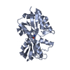
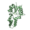
| Structure viewer | Molecule: MolmilJmol/JSmol |
|---|
- Downloads & links
-Download
| PDBx/mmCIF format | 1wdn.cif.gz | 57.4 KB | Display | PDBx/mmCIF format |
|---|---|---|---|---|
| PDB format | pdb1wdn.ent.gz | 42.2 KB | Display | PDB format |
| PDBx/mmJSON format | 1wdn.json.gz | Tree view | PDBx/mmJSON format | |
| Others |  Other downloads Other downloads |
-Validation report
| Arichive directory | https://data.pdbj.org/pub/pdb/validation_reports/wd/1wdnftp://data.pdbj.org/pub/pdb/validation_reports/wd/1wdn | HTTPS FTP |
|---|
-Related structure data
| Similar structure data |
|---|


-Links
 PDBj
PDBj
- Assembly
| Deposited unit | 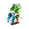
| ||||||||
|---|---|---|---|---|---|---|---|---|---|
| 1 |
| ||||||||
| Unit cell |
|
-Components
| #1: Protein | Mass: 24980.359 Da / Num. of mol.: 1 / Source method: isolated from a natural source / Details: LIGANDED CLOSE-CLEFT FORM / Source: (natural) |
|---|---|
| #2: Chemical | ChemComp-GLN /   Type: L-peptide linking / Mass: 146.144 Da / Num. of mol.: 1 / Source method: obtained synthetically / Formula: C5H10N2O3 Type: L-peptide linking / Mass: 146.144 Da / Num. of mol.: 1 / Source method: obtained synthetically / Formula: C5H10N2O3 |
| #3: Water | ChemComp-HOH /  Mass: 18.015 Da / Num. of mol.: 122 / Source method: isolated from a natural source / Formula: H2O Mass: 18.015 Da / Num. of mol.: 122 / Source method: isolated from a natural source / Formula: H2O |
-Experimental details
-Experiment
| Experiment | Method: X-RAY DIFFRACTION |
|---|
- Sample preparation
| Crystal | Density Matthews: 2.11 Å3/Da / Density % sol: 42 % | ||||||||||||||||||||||||||||||
|---|---|---|---|---|---|---|---|---|---|---|---|---|---|---|---|---|---|---|---|---|---|---|---|---|---|---|---|---|---|---|---|
| Crystal grow | *PLUS Temperature: 18 ℃ / pH: 4.6 / Method: vapor diffusion, hanging drop | ||||||||||||||||||||||||||||||
| Components of the solutions | *PLUS
|
-Data collection
| Diffraction source | Wavelength: 1.5418 |
|---|---|
| Detector | Type: RIGAKU RAXIS II / Detector: IMAGE PLATE / Date: Jan 25, 1995 |
| Radiation | Monochromatic (M) / Laue (L): M / Scattering type: x-ray |
| Radiation wavelength | Wavelength: 1.5418 Å / Relative weight: 1 |
| Reflection | Num. obs: 15110 / % possible obs: 91.1 % / Observed criterion σ(I): 0 / Redundancy: 4.5 % / Rmerge(I) obs: 0.082 |
| Reflection | *PLUS Highest resolution: 1.9 Å / Num. measured all: 65773 |
| Reflection shell | *PLUS Highest resolution: 1.94 Å / Lowest resolution: 2.03 Å / % possible obs: 82.5 % |
- Processing
| Software |
| ||||||||||||||||||||||||||||||||||||||||||||||||||||||||||||
|---|---|---|---|---|---|---|---|---|---|---|---|---|---|---|---|---|---|---|---|---|---|---|---|---|---|---|---|---|---|---|---|---|---|---|---|---|---|---|---|---|---|---|---|---|---|---|---|---|---|---|---|---|---|---|---|---|---|---|---|---|---|
| Refinement | Resolution: 1.94→6 Å / σ(F): 2 /
| ||||||||||||||||||||||||||||||||||||||||||||||||||||||||||||
| Displacement parameters | Biso mean: 24.7 Å2 | ||||||||||||||||||||||||||||||||||||||||||||||||||||||||||||
| Refine analyze | Luzzati coordinate error obs: 0.3 Å | ||||||||||||||||||||||||||||||||||||||||||||||||||||||||||||
| Refinement step | Cycle: LAST / Resolution: 1.94→6 Å
| ||||||||||||||||||||||||||||||||||||||||||||||||||||||||||||
| Refine LS restraints |
| ||||||||||||||||||||||||||||||||||||||||||||||||||||||||||||
| Software | *PLUS Name: X-PLOR / Version: 3.1 / Classification: refinement | ||||||||||||||||||||||||||||||||||||||||||||||||||||||||||||
| Refinement | *PLUS Rfactor obs: 0.2 / Rfactor Rfree: 0.3 / Rfactor Rwork: 0.2 | ||||||||||||||||||||||||||||||||||||||||||||||||||||||||||||
| Solvent computation | *PLUS | ||||||||||||||||||||||||||||||||||||||||||||||||||||||||||||
| Displacement parameters | *PLUS |
