 Movie
Movie Controller


+ Open data

- Basic information
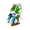
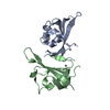
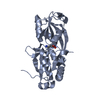
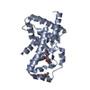
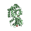
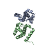
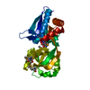
| Entry | Database: PDB / ID: 6svf | ||||||
|---|---|---|---|---|---|---|---|
| Title | Crystal structure of the P235GK mutant of ArgBP from T. maritima | ||||||
 Components Components | Amino acid ABC transporter, periplasmic amino acid-binding protein | ||||||
 Keywords Keywords | TRANSPORT PROTEIN / DOMAIN SWAPPING / MUTAGENESIS / SUBSTRATE BINDING PROTEIN | ||||||
| Function / homology |  Function and homology information Function and homology informationamino acid binding / ligand-gated monoatomic ion channel activity / outer membrane-bounded periplasmic space / membrane Similarity search - Function | ||||||
| Biological species |   Thermotoga maritima (bacteria) Thermotoga maritima (bacteria) | ||||||
| Method |  X-RAY DIFFRACTION / MOLECULAR REPLACEMENT / Resolution: 1.6 Å X-RAY DIFFRACTION / MOLECULAR REPLACEMENT / Resolution: 1.6 Å | ||||||
 Authors Authors | Vitagliano, L. / Berisio, R. / Esposito, L. / Balasco, N. / Smaldone, G. / Ruggiero, A. | ||||||
 Citation Citation | Journal: Acta Crystallogr.,Sect.F / Year: 2019 Title: The non-swapped monomeric structure of the arginine-binding protein from Thermotoga maritima. Authors: Smaldone, G. / Ruggiero, A. / Balasco, N. / Abuhammad, A. / Autiero, I. / Caruso, D. / Esposito, D. / Ferraro, G. / Gelardi, E.L.M. / Moreira, M. / Quareshy, M. / Romano, M. / Saaret, A. / ...Authors: Smaldone, G. / Ruggiero, A. / Balasco, N. / Abuhammad, A. / Autiero, I. / Caruso, D. / Esposito, D. / Ferraro, G. / Gelardi, E.L.M. / Moreira, M. / Quareshy, M. / Romano, M. / Saaret, A. / Selvam, I. / Squeglia, F. / Troisi, R. / Kroon-Batenburg, L.M.J. / Esposito, L. / Berisio, R. / Vitagliano, L. | ||||||
| History |
|
- Structure visualization
| Structure viewer | Molecule: MolmilJmol/JSmol |
|---|
- Downloads & links
-Download
| PDBx/mmCIF format | 6svf.cif.gz | 68.4 KB | Display | PDBx/mmCIF format |
|---|---|---|---|---|
| PDB format | pdb6svf.ent.gz | 49 KB | Display | PDB format |
| PDBx/mmJSON format | 6svf.json.gz | Tree view | PDBx/mmJSON format | |
| Others |  Other downloads Other downloads |
-Validation report
| Arichive directory | https://data.pdbj.org/pub/pdb/validation_reports/sv/6svfftp://data.pdbj.org/pub/pdb/validation_reports/sv/6svf | HTTPS FTP |
|---|
-Related structure data
| Related structure data | 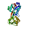 6ggpS S: Starting model for refinement |
|---|---|
| Similar structure data |


-Links
 PDBj
PDBj

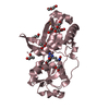
- Assembly
| Deposited unit | 
| ||||||||
|---|---|---|---|---|---|---|---|---|---|
| 1 |
| ||||||||
| Unit cell |
|
-Components
| #1: Protein | Mass: 25518.279 Da / Num. of mol.: 1 Source method: isolated from a genetically manipulated source Source: (gene. exp.) Thermotoga maritima (strain ATCC 43589 / MSB8 / DSM 3109 / JCM 10099) (bacteria)Strain: ATCC 43589 / MSB8 / DSM 3109 / JCM 10099 / Gene: TM_0593 / Production host: |
|---|---|
| #2: Chemical | ChemComp-ARG /   Type: L-peptide linking / Mass: 175.209 Da / Num. of mol.: 1 / Source method: obtained synthetically / Formula: C6H15N4O2 Type: L-peptide linking / Mass: 175.209 Da / Num. of mol.: 1 / Source method: obtained synthetically / Formula: C6H15N4O2 |
| #3: Water | ChemComp-HOH /  Mass: 18.015 Da / Num. of mol.: 355 / Source method: isolated from a natural source / Formula: H2O Mass: 18.015 Da / Num. of mol.: 355 / Source method: isolated from a natural source / Formula: H2O |
| Has ligand of interest | N |
-Experimental details
-Experiment
| Experiment | Method: X-RAY DIFFRACTION / Number of used crystals: 1 |
|---|
- Sample preparation
| Crystal | Density Matthews: 2.27 Å3/Da / Density % sol: 45.81 % |
|---|---|
| Crystal grow | Temperature: 293 K / Method: vapor diffusion, hanging drop Details: protein concentration in the range 6-10 mg/ml in a solution containing 2.0 M ammonium sulfate and 5 % (v/v) 2-propanol |
-Data collection
| Diffraction | Mean temperature: 100 K / Serial crystal experiment: N |
|---|---|
| Diffraction source | Source: ROTATING ANODE / Type: RIGAKU MICROMAX-007 HF / Wavelength: 1.54 Å |
| Detector | Type: RIGAKU SATURN 944 / Detector: CCD / Date: Sep 28, 2017 |
| Radiation | Protocol: SINGLE WAVELENGTH / Monochromatic (M) / Laue (L): M / Scattering type: x-ray |
| Radiation wavelength | Wavelength: 1.54 Å / Relative weight: 1 |
| Reflection | Resolution: 1.6→30 Å / Num. obs: 30630 / % possible obs: 97.5 % / Redundancy: 3.4 % / Rpim(I) all: 0.037 / Rrim(I) all: 0.08 / Rsym value: 0.07 / Net I/σ(I): 10.9 |
| Reflection shell | Resolution: 1.6→1.657 Å / Num. unique obs: 2798 / Rpim(I) all: 0.227 / Rrim(I) all: 0.345 / Rsym value: 0.258 |
- Processing
| Software |
| ||||||||||||||||||||||||||||||||||||||||||||||||||||||||||||
|---|---|---|---|---|---|---|---|---|---|---|---|---|---|---|---|---|---|---|---|---|---|---|---|---|---|---|---|---|---|---|---|---|---|---|---|---|---|---|---|---|---|---|---|---|---|---|---|---|---|---|---|---|---|---|---|---|---|---|---|---|---|
| Refinement | Method to determine structure: MOLECULAR REPLACEMENT Starting model: 6GGP Resolution: 1.6→30 Å / Cor.coef. Fo:Fc: 0.962 / Cor.coef. Fo:Fc free: 0.938 / SU B: 1.94 / SU ML: 0.065 / Cross valid method: THROUGHOUT / σ(F): 0 / ESU R: 0.092 / ESU R Free: 0.098 Details: HYDROGENS HAVE BEEN ADDED IN THE RIDING POSITIONS U VALUES : REFINED INDIVIDUALLY
| ||||||||||||||||||||||||||||||||||||||||||||||||||||||||||||
| Solvent computation | Ion probe radii: 0.8 Å / Shrinkage radii: 0.8 Å / VDW probe radii: 1.2 Å | ||||||||||||||||||||||||||||||||||||||||||||||||||||||||||||
| Displacement parameters | Biso max: 47.88 Å2 / Biso mean: 14.06 Å2 / Biso min: 5.3 Å2
| ||||||||||||||||||||||||||||||||||||||||||||||||||||||||||||
| Refinement step | Cycle: final / Resolution: 1.6→30 Å
| ||||||||||||||||||||||||||||||||||||||||||||||||||||||||||||
| Refine LS restraints |
| ||||||||||||||||||||||||||||||||||||||||||||||||||||||||||||
| LS refinement shell | Resolution: 1.6→1.641 Å / Rfactor Rfree error: 0 / Total num. of bins used: 20
|
