Redundancy: 6.3 % / Av σ(I) over netI: 16.2 / Number: 96529 / Rmerge(I) obs: 0.057 / Χ2: 1.02 / D res high: 2.9 Å / D res low: 20 Å / Num. obs: 15305 / % possible obs: 99.8
Diffraction reflection shell
Highest resolution (Å)
Lowest resolution (Å)
% possible obs (%)
ID
Rmerge(I) obs
Chi squared
Redundancy
6.19
20
100
1
0.036
1.019
9.3
4.94
6.19
100
1
0.054
1.036
9.2
4.32
4.94
100
1
0.05
1.004
8
3.93
4.32
100
1
0.053
1.006
6.4
3.65
3.93
99.8
1
0.059
1.019
5.2
3.44
3.65
99.9
1
0.076
1.04
5.2
3.26
3.44
99.9
1
0.112
1.026
5.2
3.12
3.26
100
1
0.162
1.007
5.1
3
3.12
99.9
1
0.241
1.035
4.9
2.9
3
98.1
1
0.283
1.036
4.4
Reflection
Resolution: 2.4→20 Å / Num. obs: 14718 / % possible obs: 100 % / Redundancy: 5.1 % / Rmerge(I) obs: 0.046 / Χ2: 1.03 / Net I/σ(I): 14.7
Reflection shell
Resolution (Å)
Redundancy (%)
Rmerge(I) obs
Num. unique all
Χ2
% possible all
2.4-2.49
4.2
0.419
1434
1.053
99.9
2.49-2.58
4.3
0.331
1438
1.053
100
2.58-2.7
4.3
0.247
1425
1.035
100
2.7-2.84
4.3
0.171
1460
1.021
100
2.84-3.02
4.3
0.116
1444
1.033
99.9
3.02-3.25
4.4
0.087
1459
1.02
99.9
3.25-3.58
4.7
0.059
1459
1.042
99.8
3.58-4.09
5.6
0.043
1492
1.022
100
4.09-5.14
7.1
0.034
1503
0.994
99.9
5.14-20
7.2
0.031
1604
1.047
100
-
Phasing
Phasing
Method: MAD
-
Processing
Software
Name
Version
Classification
NB
DENZO
datareduction
SCALEPACK
datascaling
SHARP
phasing
DM
phasing
CNS
1.1
refinement
PDB_EXTRACT
3.004
dataextraction
Refinement
Method to determine structure: MAD / Resolution: 2.4→19.87 Å / Rfactor Rfree error: 0.011 / Data cutoff high absF: 1761706.125 / Data cutoff low absF: 0 / Isotropic thermal model: RESTRAINED / Cross valid method: THROUGHOUT / σ(F): 0
In the structure databanks used in Yorodumi, some data are registered as the other names, "COVID-19 virus" and "2019-nCoV". Here are the details of the virus and the list of structure data.
Jan 31, 2019. EMDB accession codes are about to change! (news from PDBe EMDB page)
EMDB accession codes are about to change! (news from PDBe EMDB page)
The allocation of 4 digits for EMDB accession codes will soon come to an end. Whilst these codes will remain in use, new EMDB accession codes will include an additional digit and will expand incrementally as the available range of codes is exhausted. The current 4-digit format prefixed with “EMD-” (i.e. EMD-XXXX) will advance to a 5-digit format (i.e. EMD-XXXXX), and so on. It is currently estimated that the 4-digit codes will be depleted around Spring 2019, at which point the 5-digit format will come into force.
The EM Navigator/Yorodumi systems omit the EMD- prefix.
Related info.:Q: What is EMD? / ID/Accession-code notation in Yorodumi/EM Navigator
Yorodumi is a browser for structure data from EMDB, PDB, SASBDB, etc.
This page is also the successor to EM Navigator detail page, and also detail information page/front-end page for Omokage search.
The word "yorodu" (or yorozu) is an old Japanese word meaning "ten thousand". "mi" (miru) is to see.
Related info.:EMDB / PDB / SASBDB / Comparison of 3 databanks / Yorodumi Search / Aug 31, 2016. New EM Navigator & Yorodumi / Yorodumi Papers / Jmol/JSmol / Function and homology information / Changes in new EM Navigator and Yorodumi
 Movie
Movie Controller

 Yorodumi
 Open data
 Basic information
 Components
 Keywords
 Function and homology information
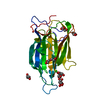
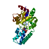
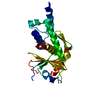
 Geobacter sulfurreducens (bacteria)
 X-RAY DIFFRACTION /
  Authors
 Citation
 Structure visualization
 Downloads & links
 Other downloads


 PDBj
PDBj

 Assembly

 Mass: 22.990 Da / Num. of mol.: 1 / Source method: obtained synthetically / Formula: Na
 Mass: 18.015 Da / Num. of mol.: 141 / Source method: isolated from a natural source / Formula: H2O
 Sample preparation
 / Beamline: X4A / Wavelength: 0.9870, 0.9795, 0.9791, 0.9715
 Processing