Mass: 18.015 Da / Num. of mol.: 271 / Source method: isolated from a natural source / Formula: H2O
-
Details
Has protein modification
Y
Nonpolymer details
B-D-GALACTOSE (GAL): BUILDS GALB1-3GLC WITH GLUCOSE. ALPHA-D-GLUCOSE (GLC): BUILDS GALB1-3GLC WITH ...B-D-GALACTOSE (GAL): BUILDS GALB1-3GLC WITH GLUCOSE. ALPHA-D-GLUCOSE (GLC): BUILDS GALB1-3GLC WITH GALACTOSE. IS A MIXTURE OF ALPHA AND BETA GLUCOSE
Sequence details
WE WORKED WITH THE N-TERMINAL ADHESIVE DOMAIN OF EPA1A, WHILE Q6VBJ0 PRESENTS THE ENTIRE SEQUENCE. ...WE WORKED WITH THE N-TERMINAL ADHESIVE DOMAIN OF EPA1A, WHILE Q6VBJ0 PRESENTS THE ENTIRE SEQUENCE. WE USED Q6VBJ0 SEQUENCE NUMBERING THROUGHOUT THE PRESENT WORK.
-
Experimental details
-
Experiment
Experiment
Method: X-RAY DIFFRACTION / Number of used crystals: 1
-
Sample preparation
Crystal
Density Matthews: 2.31 Å3/Da / Density % sol: 46.75 % / Description: NONE
Crystal grow
pH: 6.5 Details: 0.1 M MES PH 6.5, 0.2 M AMMONIUM SULFATE, 0.025 M LACTOSE
Resolution: 1.5→19.89 Å / Cor.coef. Fo:Fc: 0.969 / Cor.coef. Fo:Fc free: 0.961 / SU B: 2.557 / SU ML: 0.044 / Cross valid method: THROUGHOUT / ESU R: 0.062 / ESU R Free: 0.066 / Stereochemistry target values: MAXIMUM LIKELIHOOD / Details: HYDROGENS HAVE BEEN ADDED IN THE RIDING POSITIONS.
Rfactor
Num. reflection
% reflection
Selection details
Rfree
0.18576
1498
3.5 %
RANDOM
Rwork
0.15579
-
-
-
obs
0.15683
41751
98.46 %
-
Solvent computation
Ion probe radii: 0.8 Å / Shrinkage radii: 0.8 Å / VDW probe radii: 1.2 Å / Solvent model: BABINET MODEL WITH MASK
 Movie
Movie Controller

 Yorodumi
 Open data
 Basic information
 Components
 Keywords
 Function and homology information
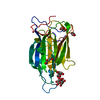
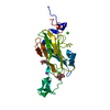
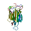
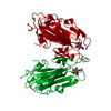
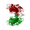
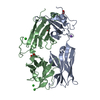
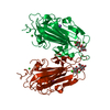
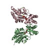
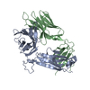
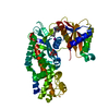
 CANDIDA GLABRATA (fungus)
 X-RAY DIFFRACTION /
  Authors
 Citation
 Structure visualization
 Downloads & links
 Other downloads
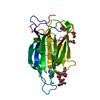


 PDBj
PDBj
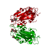
 Assembly
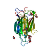


 Mass: 40.078 Da / Num. of mol.: 1 / Source method: obtained synthetically / Formula: Ca
 Mass: 92.094 Da / Num. of mol.: 3 / Source method: obtained synthetically / Formula: C3H8O3
 Mass: 106.120 Da / Num. of mol.: 1 / Source method: obtained synthetically / Formula: C4H10O3
 Sample preparation
 / Beamline: ID14-2 / Wavelength: 0.933
  Processing