 Movie
Movie Controller

[English] 日本語
 Yorodumi
- PDB-2xjp: X-ray structure of the N-terminal domain of the flocculin Flo5 fr... -
+ Open data

- Basic information
| Entry | Database: PDB / ID: 2xjp | ||||||
|---|---|---|---|---|---|---|---|
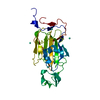
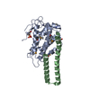
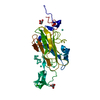
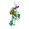
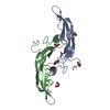
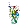
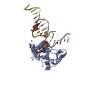
| Title | X-ray structure of the N-terminal domain of the flocculin Flo5 from Saccharomyces cerevisiae in complex with calcium and mannose | ||||||
 Components Components | FLOCCULATION PROTEIN FLO5 | ||||||
 Keywords Keywords | CELL ADHESION / GREENBEARD / PA14-DOMAIN / CARBOHYDRATE BINDING / SOCIAL INTERACTION | ||||||
| Function / homology |  Function and homology information Function and homology informationflocculation / fungal-type cell wall / cell-substrate adhesion / D-mannose binding / side of membrane / cell periphery Similarity search - Function | ||||||
| Biological species |  | ||||||
| Method |  X-RAY DIFFRACTION / SYNCHROTRON / SIRAS / Resolution: 0.95 Å X-RAY DIFFRACTION / SYNCHROTRON / SIRAS / Resolution: 0.95 Å | ||||||
 Authors Authors | Veelders, M. / Brueckner, S. / Ott, D. / Unverzagt, C. / Moesch, H.-U. / Essen, L.-O. | ||||||
 Citation Citation | Journal: Proc.Natl.Acad.Sci.USA / Year: 2010 Title: Structural Basis of Flocculin-Mediated Social Behavior in Yeast Authors: Veelders, M. / Brueckner, S. / Ott, D. / Unverzagt, C. / Moesch, H.-U. / Essen, L.-O. | ||||||
| History |
|
- Structure visualization
| Structure viewer | Molecule: MolmilJmol/JSmol |
|---|
- Downloads & links
-Download
| PDBx/mmCIF format | 2xjp.cif.gz | 242.6 KB | Display | PDBx/mmCIF format |
|---|---|---|---|---|
| PDB format | pdb2xjp.ent.gz | 200.4 KB | Display | PDB format |
| PDBx/mmJSON format | 2xjp.json.gz | Tree view | PDBx/mmJSON format | |
| Others |  Other downloads Other downloads |
-Validation report
| Arichive directory | https://data.pdbj.org/pub/pdb/validation_reports/xj/2xjpftp://data.pdbj.org/pub/pdb/validation_reports/xj/2xjp | HTTPS FTP |
|---|
-Related structure data
| Related structure data |  2xjqC  2xjrC  2xjsC  2xjtC  2xjuC 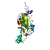 2xjvC C: citing same article ( |
|---|---|
| Similar structure data |


-Links
 PDBj
PDBj


- Assembly
| Deposited unit | 
| ||||||||
|---|---|---|---|---|---|---|---|---|---|
| 1 |
| ||||||||
| Unit cell |
|
-Components
-Protein , 1 types, 1 molecules A
| #1: Protein | Mass: 27732.512 Da / Num. of mol.: 1 / Fragment: LECTIN-LIKE FLO5A-DOMAIN, RESIDUES 23-271 Source method: isolated from a genetically manipulated source Source: (gene. exp.) Strain: S288C / Production host:  |
|---|
-Sugars , 2 types, 3 molecules 

| #5: Sugar | ChemComp-MAN /  Type: D-saccharide, alpha linking / Mass: 180.156 Da / Num. of mol.: 1 Type: D-saccharide, alpha linking / Mass: 180.156 Da / Num. of mol.: 1Source method: isolated from a genetically manipulated source Formula: C6H12O6 |
|---|---|
| #6: Sugar |  Type: D-saccharide, beta linking / Mass: 180.156 Da / Num. of mol.: 2 Type: D-saccharide, beta linking / Mass: 180.156 Da / Num. of mol.: 2Source method: isolated from a genetically manipulated source Formula: C6H12O6 |
-Non-polymers , 4 types, 465 molecules 


| #2: Chemical |  Mass: 40.078 Da / Num. of mol.: 2 / Source method: obtained synthetically / Formula: Ca Mass: 40.078 Da / Num. of mol.: 2 / Source method: obtained synthetically / Formula: Ca#3: Chemical |  Mass: 22.990 Da / Num. of mol.: 2 / Source method: obtained synthetically / Formula: Na Mass: 22.990 Da / Num. of mol.: 2 / Source method: obtained synthetically / Formula: Na#4: Chemical | ChemComp-CL / |  Mass: 35.453 Da / Num. of mol.: 1 / Source method: obtained synthetically / Formula: Cl Mass: 35.453 Da / Num. of mol.: 1 / Source method: obtained synthetically / Formula: Cl#7: Water | ChemComp-HOH / | Mass: 18.015 Da / Num. of mol.: 460 / Source method: isolated from a natural source / Formula: H2O |
|---|
-Details
| Has protein modification | Y |
|---|---|
| Nonpolymer details | D-MANNOSE(MAN): MANNOSE HAS BEEN FOUND IN BOTH ANOMERIC CONFORMATION AS ACCOUNTED BY THE ...D-MANNOSE(MAN): MANNOSE HAS BEEN FOUND IN BOTH ANOMERIC CONFORMATI |
-Experimental details
-Experiment
| Experiment | Method: X-RAY DIFFRACTION / Number of used crystals: 1 |
|---|
- Sample preparation
| Crystal | Density Matthews: 2.75 Å3/Da / Density % sol: 55.27 % / Description: NONE |
|---|---|
| Crystal grow | Details: 0.5 M NACL, 0.1 M BISTRIS PH7.5, 20% PEG 4000 |
-Data collection
| Diffraction | Mean temperature: 100 K |
|---|---|
| Diffraction source | Source: SYNCHROTRON / Site: ESRF  / Beamline: ID29 / Wavelength: 0.9149 / Beamline: ID29 / Wavelength: 0.9149 |
| Detector | Type: ADSC QUANTUM 315r / Detector: CCD / Date: Nov 24, 2008 / Details: CYLINDRICAL GRAZING INCIDENCE MIRROR |
| Radiation | Monochromator: SI (311) / Protocol: SINGLE WAVELENGTH / Monochromatic (M) / Laue (L): M / Scattering type: x-ray |
| Radiation wavelength | Wavelength: 0.9149 Å / Relative weight: 1 |
| Reflection | Resolution: 0.95→40.29 Å / Num. obs: 190329 / % possible obs: 99.3 % / Observed criterion σ(I): -3 / Redundancy: 4.2 % / Biso Wilson estimate: 7 Å2 / Rmerge(I) obs: 0.03 / Net I/σ(I): 19.6 |
| Reflection shell | Resolution: 0.95→1 Å / Redundancy: 3.7 % / Rmerge(I) obs: 0.44 / Mean I/σ(I) obs: 2.8 / % possible all: 95.7 |
- Processing
| Software |
| ||||||||||||||||||||||||||||||||||||||||||||||||||||||||||||||||||||||||||||||||||||||||||||||||||||||||||||||||||||||||||||||||||||||||||||||||||||||||||||||||||||||||||||||||||||||
|---|---|---|---|---|---|---|---|---|---|---|---|---|---|---|---|---|---|---|---|---|---|---|---|---|---|---|---|---|---|---|---|---|---|---|---|---|---|---|---|---|---|---|---|---|---|---|---|---|---|---|---|---|---|---|---|---|---|---|---|---|---|---|---|---|---|---|---|---|---|---|---|---|---|---|---|---|---|---|---|---|---|---|---|---|---|---|---|---|---|---|---|---|---|---|---|---|---|---|---|---|---|---|---|---|---|---|---|---|---|---|---|---|---|---|---|---|---|---|---|---|---|---|---|---|---|---|---|---|---|---|---|---|---|---|---|---|---|---|---|---|---|---|---|---|---|---|---|---|---|---|---|---|---|---|---|---|---|---|---|---|---|---|---|---|---|---|---|---|---|---|---|---|---|---|---|---|---|---|---|---|---|---|---|
| Refinement | Method to determine structure: SIRAS Starting model: NONE Resolution: 0.95→20 Å / Cor.coef. Fo:Fc: 0.986 / Cor.coef. Fo:Fc free: 0.981 / SU B: 0.375 / SU ML: 0.009 / Cross valid method: THROUGHOUT / ESU R: 0.014 / ESU R Free: 0.015 / Stereochemistry target values: MAXIMUM LIKELIHOOD Details: HYDROGENS HAVE BEEN ADDED IN THE RIDING POSITIONS. RESIDUES 197 - 199 ARE NOT WELL DEFINED. RESIDUES 200 - 204 HAVE BEEN MODELLED IN TWO DISTINCT CONFORMATIONS.
| ||||||||||||||||||||||||||||||||||||||||||||||||||||||||||||||||||||||||||||||||||||||||||||||||||||||||||||||||||||||||||||||||||||||||||||||||||||||||||||||||||||||||||||||||||||||
| Solvent computation | Ion probe radii: 0.8 Å / Shrinkage radii: 0.8 Å / VDW probe radii: 1.2 Å / Solvent model: BABINET MODEL WITH MASK | ||||||||||||||||||||||||||||||||||||||||||||||||||||||||||||||||||||||||||||||||||||||||||||||||||||||||||||||||||||||||||||||||||||||||||||||||||||||||||||||||||||||||||||||||||||||
| Displacement parameters | Biso mean: 12.246 Å2
| ||||||||||||||||||||||||||||||||||||||||||||||||||||||||||||||||||||||||||||||||||||||||||||||||||||||||||||||||||||||||||||||||||||||||||||||||||||||||||||||||||||||||||||||||||||||
| Refinement step | Cycle: LAST / Resolution: 0.95→20 Å
| ||||||||||||||||||||||||||||||||||||||||||||||||||||||||||||||||||||||||||||||||||||||||||||||||||||||||||||||||||||||||||||||||||||||||||||||||||||||||||||||||||||||||||||||||||||||
| Refine LS restraints |
|