 Movie
Movie Controller

[English] 日本語
 Yorodumi
- PDB-6lhr: Crystal structure of the complex between Vesicle Amine Transport-... -
+ Open data

- Basic information
| Entry | Database: PDB / ID: 6lhr | ||||||
|---|---|---|---|---|---|---|---|
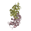
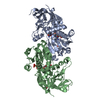
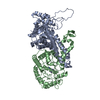
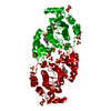
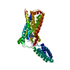
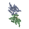
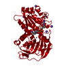
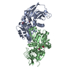
| Title | Crystal structure of the complex between Vesicle Amine Transport-1 and NADP | ||||||
 Components Components | Synaptic vesicle membrane protein VAT-1 homolog | ||||||
 Keywords Keywords | OXIDOREDUCTASE / VAT1 / Complex / Quinone | ||||||
| Function / homology |  Function and homology information Function and homology informationNADPH:quinone reductase / phospholipid transfer activity / intermembrane phospholipid transfer / NADPH dehydrogenase (quinone) activity / negative regulation of mitochondrial fusion / azurophil granule lumen / mitochondrial outer membrane / Neutrophil degranulation / extracellular exosome / extracellular region ...NADPH:quinone reductase / phospholipid transfer activity / intermembrane phospholipid transfer / NADPH dehydrogenase (quinone) activity / negative regulation of mitochondrial fusion / azurophil granule lumen / mitochondrial outer membrane / Neutrophil degranulation / extracellular exosome / extracellular region / zinc ion binding / cytoplasm / cytosol Similarity search - Function | ||||||
| Biological species |  Homo sapiens (human) Homo sapiens (human) | ||||||
| Method |  X-RAY DIFFRACTION / SYNCHROTRON / MOLECULAR REPLACEMENT / Resolution: 2.62 Å X-RAY DIFFRACTION / SYNCHROTRON / MOLECULAR REPLACEMENT / Resolution: 2.62 Å | ||||||
 Authors Authors | Hakoshima, T. / Kim, S.-Y. / Mori, T. | ||||||
 Citation Citation | Journal: Sci Rep / Year: 2021 Title: Structural insights into vesicle amine transport-1 (VAT-1) as a member of the NADPH-dependent quinone oxidoreductase family. Authors: Kim, S.-Y. / Mori, T. / Chek, M.F. / Furuya, S. / Matsumoto, K. / Yajima, T. / Ogura, T. / Hakoshima, T. | ||||||
| History |
|
- Structure visualization
| Structure viewer | Molecule: MolmilJmol/JSmol |
|---|
- Downloads & links
-Download
| PDBx/mmCIF format | 6lhr.cif.gz | 328.7 KB | Display | PDBx/mmCIF format |
|---|---|---|---|---|
| PDB format | pdb6lhr.ent.gz | 216.8 KB | Display | PDB format |
| PDBx/mmJSON format | 6lhr.json.gz | Tree view | PDBx/mmJSON format | |
| Others |  Other downloads Other downloads |
-Validation report
| Arichive directory | https://data.pdbj.org/pub/pdb/validation_reports/lh/6lhrftp://data.pdbj.org/pub/pdb/validation_reports/lh/6lhr | HTTPS FTP |
|---|
-Related structure data
| Related structure data |  6liiC 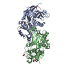 4a27S S: Starting model for refinement C: citing same article ( |
|---|---|
| Similar structure data |


-Links
 PDBj
PDBj

- Assembly
| Deposited unit | 
| ||||||||||||||||||||||||||||||||||||||||||||||||||||||||||||||||||||||||||||||||||||||||||
|---|---|---|---|---|---|---|---|---|---|---|---|---|---|---|---|---|---|---|---|---|---|---|---|---|---|---|---|---|---|---|---|---|---|---|---|---|---|---|---|---|---|---|---|---|---|---|---|---|---|---|---|---|---|---|---|---|---|---|---|---|---|---|---|---|---|---|---|---|---|---|---|---|---|---|---|---|---|---|---|---|---|---|---|---|---|---|---|---|---|---|---|
| 1 | 
| ||||||||||||||||||||||||||||||||||||||||||||||||||||||||||||||||||||||||||||||||||||||||||
| 2 | 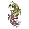
| ||||||||||||||||||||||||||||||||||||||||||||||||||||||||||||||||||||||||||||||||||||||||||
| Unit cell |
| ||||||||||||||||||||||||||||||||||||||||||||||||||||||||||||||||||||||||||||||||||||||||||
| Noncrystallographic symmetry (NCS) | NCS domain:
NCS domain segments: Ens-ID: 1
|