 Movie
Movie Controller

[English] 日本語
 Yorodumi
- PDB-2b5w: Crystal structure of D38C glucose dehydrogenase mutant from Halof... -
+ Open data

- Basic information
| Entry | Database: PDB / ID: 2b5w | ||||||
|---|---|---|---|---|---|---|---|
| Title | Crystal structure of D38C glucose dehydrogenase mutant from Haloferax mediterranei | ||||||
 Components Components | glucose dehydrogenase | ||||||
 Keywords Keywords | OXIDOREDUCTASE / Nucleotide binding motif | ||||||
| Function / homology |  Function and homology information Function and homology informationnon-phosphorylated glucose catabolic process / glucose 1-dehydrogenase [NAD(P)+] activity / glucose 1-dehydrogenase (NAD+) activity / glucose 1-dehydrogenase (NADP+) activity / glucose 1-dehydrogenase [NAD(P)+] / D-glucose binding / NADP+ binding / NAD+ binding / protein homodimerization activity / zinc ion binding / identical protein binding Similarity search - Function | ||||||
| Biological species |  Haloferax mediterranei (archaea) Haloferax mediterranei (archaea) | ||||||
| Method |  X-RAY DIFFRACTION / SYNCHROTRON / MOLECULAR REPLACEMENT / Resolution: 1.6 Å X-RAY DIFFRACTION / SYNCHROTRON / MOLECULAR REPLACEMENT / Resolution: 1.6 Å | ||||||
 Authors Authors | Britton, K.L. / Baker, P.J. / Fisher, M. / Ruzheinikov, S. / Gilmour, D.J. / Bonete, M.-J. / Ferrer, J. / Pire, C. / Esclapez, J. / Rice, D.W. | ||||||
 Citation Citation | Journal: Proc.Natl.Acad.Sci.Usa / Year: 2006 Title: Analysis of protein solvent interactions in glucose dehydrogenase from the extreme halophile Haloferax mediterranei. Authors: Britton, K.L. / Baker, P.J. / Fisher, M. / Ruzheinikov, S. / Gilmour, D.J. / Bonete, M.-J. / Ferrer, J. / Pire, C. / Esclapez, J. / Rice, D.W. #1: Journal: Acta Crystallogr.,Sect.D / Year: 2001 Title: Crystallization and Preliminary X-Ray Analysis of Glucose Dehydrogenase from Haloferax Mediterranei Authors: Ferrer, J. / Fisher, M. / Burke, J. / Sedelnikova, S.E. / Baker, P.J. / Gilmour, D.J. / Bonete, M.-J. / Pire, C. / Esclapez, J. / Rice, D.W. | ||||||
| History |
|
- Structure visualization
| Structure viewer | Molecule: MolmilJmol/JSmol |
|---|
- Downloads & links
-Download
| PDBx/mmCIF format | 2b5w.cif.gz | 107.2 KB | Display | PDBx/mmCIF format |
|---|---|---|---|---|
| PDB format | pdb2b5w.ent.gz | 78.2 KB | Display | PDB format |
| PDBx/mmJSON format | 2b5w.json.gz | Tree view | PDBx/mmJSON format | |
| Others |  Other downloads Other downloads |
-Validation report
| Arichive directory | https://data.pdbj.org/pub/pdb/validation_reports/b5/2b5wftp://data.pdbj.org/pub/pdb/validation_reports/b5/2b5w | HTTPS FTP |
|---|
-Related structure data
| Related structure data |  2b5vSC S: Starting model for refinement C: citing same article ( |
|---|---|
| Similar structure data |


-Links
 PDBj
PDBj


- Assembly
| Deposited unit | 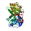
| ||||||||
|---|---|---|---|---|---|---|---|---|---|
| 1 | 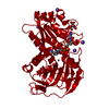
| ||||||||
| Unit cell |
| ||||||||
| Components on special symmetry positions |
|
-Components
-Protein , 1 types, 1 molecules A
| #1: Protein | Mass: 39270.781 Da / Num. of mol.: 1 / Mutation: C38D Source method: isolated from a genetically manipulated source Source: (gene. exp.) Haloferax mediterranei (archaea) / Gene: gdh / Plasmid: PET3A / Species (production host): Escherichia coli / Production host:  References: UniProt: Q977U7, glucose 1-dehydrogenase [NAD(P)+] |
|---|
-Non-polymers , 5 types, 683 molecules 


| #2: Chemical | ChemComp-ZN /  Mass: 65.409 Da / Num. of mol.: 1 / Source method: obtained synthetically / Formula: Zn Mass: 65.409 Da / Num. of mol.: 1 / Source method: obtained synthetically / Formula: Zn | ||||
|---|---|---|---|---|---|
| #3: Chemical | ChemComp-FLC /  Mass: 189.100 Da / Num. of mol.: 1 / Source method: obtained synthetically / Formula: C6H5O7 Mass: 189.100 Da / Num. of mol.: 1 / Source method: obtained synthetically / Formula: C6H5O7 | ||||
| #4: Chemical | ChemComp-K /  Mass: 39.098 Da / Num. of mol.: 5 / Source method: obtained synthetically / Formula: K Mass: 39.098 Da / Num. of mol.: 5 / Source method: obtained synthetically / Formula: K#5: Chemical | ChemComp-NAP / |  Mass: 743.405 Da / Num. of mol.: 1 / Source method: obtained synthetically / Formula: C21H28N7O17P3 Mass: 743.405 Da / Num. of mol.: 1 / Source method: obtained synthetically / Formula: C21H28N7O17P3#6: Water | ChemComp-HOH / | Mass: 18.015 Da / Num. of mol.: 675 / Source method: isolated from a natural source / Formula: H2O |
-Experimental details
-Experiment
| Experiment | Method: X-RAY DIFFRACTION / Number of used crystals: 1 |
|---|
- Sample preparation
| Crystal | Density Matthews: 3.2 Å3/Da / Density % sol: 61.51 % |
|---|---|
| Crystal grow | Temperature: 290 K / Method: vapor diffusion, hanging drop / pH: 7 Details: pH 7.00, temperature 290K, Vapor diffusion, hanging drop, VAPOR DIFFUSION, HANGING DROP, temperature 290 KK |
-Data collection
| Diffraction | Mean temperature: 100 K |
|---|---|
| Diffraction source | Source: SYNCHROTRON / Site: SRS  / Beamline: PX14.2 / Wavelength: 0.97 / Beamline: PX14.2 / Wavelength: 0.97 |
| Detector | Type: ADSC QUANTUM 4 / Detector: CCD / Date: Jan 16, 2002 / Details: MIRRORS |
| Radiation | Monochromator: SI 111 / Protocol: SINGLE WAVELENGTH / Monochromatic (M) / Laue (L): M / Scattering type: x-ray |
| Radiation wavelength | Wavelength: 0.97 Å / Relative weight: 1 |
| Reflection | Resolution: 1.6→20 Å / Num. obs: 66155 / % possible obs: 99.1 % / Observed criterion σ(I): 0 / Redundancy: 5.1 % / Rmerge(I) obs: 0.058 / Net I/σ(I): 25.85 |
| Reflection shell | Resolution: 1.6→1.64 Å / Rmerge(I) obs: 0.543 / Mean I/σ(I) obs: 2.69 / % possible all: 98.2 |
- Processing
| Software |
| ||||||||||||||||||||||||||||||||||||||||||||||||||||||||||||||||||||||||||||||||||||||||||||||||||||||||||||||||||||||||||||||||||||||||||||||||||||||||||||||||||||||||||
|---|---|---|---|---|---|---|---|---|---|---|---|---|---|---|---|---|---|---|---|---|---|---|---|---|---|---|---|---|---|---|---|---|---|---|---|---|---|---|---|---|---|---|---|---|---|---|---|---|---|---|---|---|---|---|---|---|---|---|---|---|---|---|---|---|---|---|---|---|---|---|---|---|---|---|---|---|---|---|---|---|---|---|---|---|---|---|---|---|---|---|---|---|---|---|---|---|---|---|---|---|---|---|---|---|---|---|---|---|---|---|---|---|---|---|---|---|---|---|---|---|---|---|---|---|---|---|---|---|---|---|---|---|---|---|---|---|---|---|---|---|---|---|---|---|---|---|---|---|---|---|---|---|---|---|---|---|---|---|---|---|---|---|---|---|---|---|---|---|---|---|---|
| Refinement | Method to determine structure: MOLECULAR REPLACEMENT Starting model: PDB ENTRY 2B5V Resolution: 1.6→20 Å / Cor.coef. Fo:Fc: 0.975 / Cor.coef. Fo:Fc free: 0.966 / SU B: 1.374 / SU ML: 0.048 / Cross valid method: THROUGHOUT / σ(F): 0 / ESU R: 0.068 / ESU R Free: 0.072 / Stereochemistry target values: Engh & Huber
| ||||||||||||||||||||||||||||||||||||||||||||||||||||||||||||||||||||||||||||||||||||||||||||||||||||||||||||||||||||||||||||||||||||||||||||||||||||||||||||||||||||||||||
| Solvent computation | Ion probe radii: 0.8 Å / Shrinkage radii: 0.8 Å / VDW probe radii: 1.4 Å / Solvent model: BABINET MODEL WITH MASK | ||||||||||||||||||||||||||||||||||||||||||||||||||||||||||||||||||||||||||||||||||||||||||||||||||||||||||||||||||||||||||||||||||||||||||||||||||||||||||||||||||||||||||
| Displacement parameters | Biso mean: 21.16 Å2 | ||||||||||||||||||||||||||||||||||||||||||||||||||||||||||||||||||||||||||||||||||||||||||||||||||||||||||||||||||||||||||||||||||||||||||||||||||||||||||||||||||||||||||
| Refinement step | Cycle: LAST / Resolution: 1.6→20 Å
| ||||||||||||||||||||||||||||||||||||||||||||||||||||||||||||||||||||||||||||||||||||||||||||||||||||||||||||||||||||||||||||||||||||||||||||||||||||||||||||||||||||||||||
| Refine LS restraints |
| ||||||||||||||||||||||||||||||||||||||||||||||||||||||||||||||||||||||||||||||||||||||||||||||||||||||||||||||||||||||||||||||||||||||||||||||||||||||||||||||||||||||||||
| LS refinement shell | Resolution: 1.6→1.64 Å / Total num. of bins used: 20 /
|
