 Movie
Movie Controller

[English] 日本語
 Yorodumi
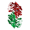
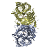
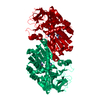
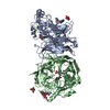
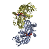
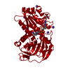
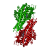
- PDB-2vwh: Haloferax mediterranei glucose dehydrogenase in complex with NADP... -
+ Open data

- Basic information
| Entry | Database: PDB / ID: 2vwh | ||||||
|---|---|---|---|---|---|---|---|
| Title | Haloferax mediterranei glucose dehydrogenase in complex with NADP, Zn and glucose. | ||||||
 Components Components | GLUCOSE DEHYDROGENASE | ||||||
 Keywords Keywords | OXIDOREDUCTASE / GLUCOSE DEHYDROGENASE / ZINC DEPENDENT MEDIUM CHAIN ALCOHOL DEHYDROGENASE FAMILY / ALCOHOL DEHYDROGENASE | ||||||
| Function / homology |  Function and homology information Function and homology informationnon-phosphorylated glucose catabolic process / glucose 1-dehydrogenase [NAD(P)+] activity / glucose 1-dehydrogenase (NAD+) activity / glucose 1-dehydrogenase (NADP+) activity / glucose 1-dehydrogenase [NAD(P)+] / D-glucose binding / NADP+ binding / NAD+ binding / protein homodimerization activity / zinc ion binding / identical protein binding Similarity search - Function | ||||||
| Biological species |  HALOFERAX MEDITERRANEI (archaea) HALOFERAX MEDITERRANEI (archaea) | ||||||
| Method |  X-RAY DIFFRACTION / MOLECULAR REPLACEMENT / Resolution: 2.03 Å X-RAY DIFFRACTION / MOLECULAR REPLACEMENT / Resolution: 2.03 Å | ||||||
 Authors Authors | Baker, P.J. / Britton, K.L. / Fisher, M. / Esclapez, J. / Pire, C. / Bonete, M.J. / Ferrer, J. / Rice, D.W. | ||||||
 Citation Citation | Journal: Proc. Natl. Acad. Sci. U.S.A. / Year: 2009 Title: Active site dynamics in the zinc-dependent medium chain alcohol dehydrogenase superfamily. Authors: Baker, P.J. / Britton, K.L. / Fisher, M. / Esclapez, J. / Pire, C. / Bonete, M.J. / Ferrer, J. / Rice, D.W. | ||||||
| History |
| ||||||
| Remark 700 | SHEET THE SHEET STRUCTURE OF THIS MOLECULE IS BIFURCATED. IN ORDER TO REPRESENT THIS FEATURE IN ... SHEET THE SHEET STRUCTURE OF THIS MOLECULE IS BIFURCATED. IN ORDER TO REPRESENT THIS FEATURE IN THE SHEET RECORDS BELOW, TWO SHEETS ARE DEFINED. |
- Structure visualization
| Structure viewer | Molecule: MolmilJmol/JSmol |
|---|
- Downloads & links
-Download
| PDBx/mmCIF format | 2vwh.cif.gz | 89.2 KB | Display | PDBx/mmCIF format |
|---|---|---|---|---|
| PDB format | pdb2vwh.ent.gz | 65.4 KB | Display | PDB format |
| PDBx/mmJSON format | 2vwh.json.gz | Tree view | PDBx/mmJSON format | |
| Others |  Other downloads Other downloads |
-Validation report
| Arichive directory | https://data.pdbj.org/pub/pdb/validation_reports/vw/2vwhftp://data.pdbj.org/pub/pdb/validation_reports/vw/2vwh | HTTPS FTP |
|---|
-Related structure data
| Related structure data |  2vwgC 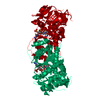 2vwpC  2vwqC  2b5vS C: citing same article ( S: Starting model for refinement |
|---|---|
| Similar structure data |


-Links
 PDBj
PDBj


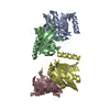
- Assembly
| Deposited unit | 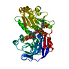
| ||||||||
|---|---|---|---|---|---|---|---|---|---|
| 1 | 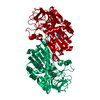
| ||||||||
| Unit cell |
| ||||||||
| Components on special symmetry positions |
|
-Components
| #1: Protein | Mass: 39282.727 Da / Num. of mol.: 1 Source method: isolated from a genetically manipulated source Source: (gene. exp.) HALOFERAX MEDITERRANEI (archaea) / Plasmid: PET3A / Production host:  References: UniProt: Q977U7, glucose 1-dehydrogenase [NAD(P)+] |
|---|---|
| #2: Chemical | ChemComp-NAP /   Mass: 743.405 Da / Num. of mol.: 1 / Source method: obtained synthetically / Formula: C21H28N7O17P3 Mass: 743.405 Da / Num. of mol.: 1 / Source method: obtained synthetically / Formula: C21H28N7O17P3 |
| #3: Sugar | ChemComp-BGC /   Type: D-saccharide, beta linking / Mass: 180.156 Da / Num. of mol.: 1 Type: D-saccharide, beta linking / Mass: 180.156 Da / Num. of mol.: 1Source method: isolated from a genetically manipulated source Formula: C6H12O6 |
| #4: Chemical | ChemComp-ZN /   Mass: 65.409 Da / Num. of mol.: 1 / Source method: obtained synthetically / Formula: Zn Mass: 65.409 Da / Num. of mol.: 1 / Source method: obtained synthetically / Formula: Zn |
| #5: Water | ChemComp-HOH /  Mass: 18.015 Da / Num. of mol.: 148 / Source method: isolated from a natural source / Formula: H2O Mass: 18.015 Da / Num. of mol.: 148 / Source method: isolated from a natural source / Formula: H2O |
-Experimental details
-Experiment
| Experiment | Method: X-RAY DIFFRACTION / Number of used crystals: 1 |
|---|
- Sample preparation
| Crystal | Density Matthews: 3.3 Å3/Da / Density % sol: 63 % / Description: NONE |
|---|---|
| Crystal grow | pH: 7.5 Details: 1.5M SODIUM CITRATE, 0.1M SODIUM HEPES PH8.0, 10MM NADP, pH 7.5 |
-Data collection
| Diffraction | Mean temperature: 290 K |
|---|---|
| Diffraction source | Source: ROTATING ANODE / Type: RIGAKU RU200 / Wavelength: 1.5418 |
| Detector | Type: MARRESEARCH / Detector: IMAGE PLATE / Date: Jan 12, 2001 / Details: MIRRORS |
| Radiation | Monochromator: NI FILTER / Protocol: SINGLE WAVELENGTH / Monochromatic (M) / Laue (L): M / Scattering type: x-ray |
| Radiation wavelength | Wavelength: 1.5418 Å / Relative weight: 1 |
| Reflection | Resolution: 2.03→30 Å / Num. obs: 34033 / % possible obs: 98.9 % / Observed criterion σ(I): 0 / Redundancy: 6.8 % / Rmerge(I) obs: 0.08 / Net I/σ(I): 18.3 |
| Reflection shell | Resolution: 2.03→2.08 Å / Redundancy: 2.3 % / Rmerge(I) obs: 0.28 / Mean I/σ(I) obs: 3.7 / % possible all: 89.9 |
- Processing
| Software |
| ||||||||||||||||||||||||||||||||||||||||||||||||||||||||||||||||||||||||||||||||||||||||||||||||||||||||||||||||||||||||||||||||||||||||||||||||||||||||||||||||||||||||||||||||||||||
|---|---|---|---|---|---|---|---|---|---|---|---|---|---|---|---|---|---|---|---|---|---|---|---|---|---|---|---|---|---|---|---|---|---|---|---|---|---|---|---|---|---|---|---|---|---|---|---|---|---|---|---|---|---|---|---|---|---|---|---|---|---|---|---|---|---|---|---|---|---|---|---|---|---|---|---|---|---|---|---|---|---|---|---|---|---|---|---|---|---|---|---|---|---|---|---|---|---|---|---|---|---|---|---|---|---|---|---|---|---|---|---|---|---|---|---|---|---|---|---|---|---|---|---|---|---|---|---|---|---|---|---|---|---|---|---|---|---|---|---|---|---|---|---|---|---|---|---|---|---|---|---|---|---|---|---|---|---|---|---|---|---|---|---|---|---|---|---|---|---|---|---|---|---|---|---|---|---|---|---|---|---|---|---|
| Refinement | Method to determine structure: MOLECULAR REPLACEMENT Starting model: PDB ENTRY 2B5V Resolution: 2.03→14.87 Å / Cor.coef. Fo:Fc: 0.975 / Cor.coef. Fo:Fc free: 0.968 / SU B: 2.405 / SU ML: 0.066 / Cross valid method: THROUGHOUT / ESU R: 0.107 / ESU R Free: 0.1 / Stereochemistry target values: MAXIMUM LIKELIHOOD / Details: HYDROGENS HAVE BEEN ADDED IN THE RIDING POSITIONS.
| ||||||||||||||||||||||||||||||||||||||||||||||||||||||||||||||||||||||||||||||||||||||||||||||||||||||||||||||||||||||||||||||||||||||||||||||||||||||||||||||||||||||||||||||||||||||
| Solvent computation | Ion probe radii: 0.8 Å / Shrinkage radii: 0.8 Å / VDW probe radii: 1.4 Å / Solvent model: BABINET MODEL WITH MASK | ||||||||||||||||||||||||||||||||||||||||||||||||||||||||||||||||||||||||||||||||||||||||||||||||||||||||||||||||||||||||||||||||||||||||||||||||||||||||||||||||||||||||||||||||||||||
| Displacement parameters | Biso mean: 29.38 Å2
| ||||||||||||||||||||||||||||||||||||||||||||||||||||||||||||||||||||||||||||||||||||||||||||||||||||||||||||||||||||||||||||||||||||||||||||||||||||||||||||||||||||||||||||||||||||||
| Refinement step | Cycle: LAST / Resolution: 2.03→14.87 Å
| ||||||||||||||||||||||||||||||||||||||||||||||||||||||||||||||||||||||||||||||||||||||||||||||||||||||||||||||||||||||||||||||||||||||||||||||||||||||||||||||||||||||||||||||||||||||
| Refine LS restraints |
|