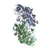
Entry Database : PDB / ID : 4a27Title Crystal structure of human synaptic vesicle membrane protein VAT-1 homolog-like protein SYNAPTIC VESICLE MEMBRANE PROTEIN VAT-1 HOMOLOG-LIKE Keywords Function / homology Function Domain/homology Component
/ / / / / / / / / / / / / / / / / / / / / / / / / Biological species HOMO SAPIENS (human)Method / / / Resolution : 2.1 Å Authors Vollmar, M. / Shafqat, N. / Muniz, J.R.C. / Krojer, T. / Allerston, C. / von Delft, F. / Bountra, C. / Arrowsmith, C.H. / Weigelt, J. / Edwards, A. ...Vollmar, M. / Shafqat, N. / Muniz, J.R.C. / Krojer, T. / Allerston, C. / von Delft, F. / Bountra, C. / Arrowsmith, C.H. / Weigelt, J. / Edwards, A. / Yue, W.W. / Oppermann, U. Journal : To be Published Title : Crystal Structure of Human Synaptic Vesicle Membrane Protein Vat-1 Homolog-Like ProteinAuthors : Vollmar, M. / Shafqat, N. / Muniz, J.R.C. / Krojer, T. / Allerston, C. / von Delft, F. / Bountra, C. / Arrowsmith, C.H. / Weigelt, J. / Edwards, A. / Yue, W.W. / Oppermann, U. History Deposition Sep 22, 2011 Deposition site / Processing site Revision 1.0 Oct 19, 2011 Provider / Type Revision 1.1 Nov 14, 2012 Group Revision 1.2 Jan 24, 2018 Group / Category / Item Revision 1.3 Dec 20, 2023 Group Data collection / Database references ... Data collection / Database references / Derived calculations / Other / Refinement description Category chem_comp_atom / chem_comp_bond ... chem_comp_atom / chem_comp_bond / database_2 / pdbx_database_status / pdbx_initial_refinement_model / struct_site Item _database_2.pdbx_DOI / _database_2.pdbx_database_accession ... _database_2.pdbx_DOI / _database_2.pdbx_database_accession / _pdbx_database_status.status_code_sf / _struct_site.pdbx_auth_asym_id / _struct_site.pdbx_auth_comp_id / _struct_site.pdbx_auth_seq_id
Show all Show less
 Movie
Movie Controller

 Yorodumi
 Open data
 Basic information
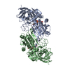
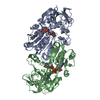
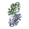
 Components
 Keywords
 Function and homology information
 HOMO SAPIENS (human)
 X-RAY DIFFRACTION /
  Authors
 Citation
 Structure visualization
 Downloads & links
 Other downloads

 PDBj
PDBj

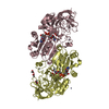
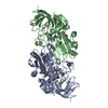
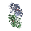
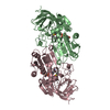
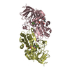
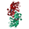
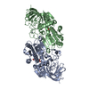
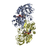
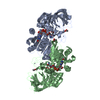
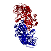
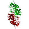
 Assembly
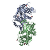


 Mass: 62.068 Da / Num. of mol.: 9 / Source method: obtained synthetically / Formula: C2H6O2
 Mass: 18.015 Da / Num. of mol.: 137 / Source method: isolated from a natural source / Formula: H2O
 Sample preparation
 / Beamline: I02 / Wavelength: 0.97
  Processing