 Movie
Movie Controller

[English] 日本語
 Yorodumi
- PDB-5kj1: G173A horse liver alcohol dehydrogenase complexed with NAD+ and p... -
+ Open data

- Basic information
| Entry | Database: PDB / ID: 5kj1 | ||||||
|---|---|---|---|---|---|---|---|
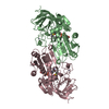
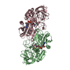
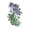
| Title | G173A horse liver alcohol dehydrogenase complexed with NAD+ and pentafluorobenzyl alcohol | ||||||
 Components Components | Alcohol dehydrogenase E chain | ||||||
 Keywords Keywords | OXIDOREDUCTASE / OXIDOREDUCTASE Inhibitor complex Rossmann fold dynamics | ||||||
| Function / homology |  Function and homology information Function and homology informationall-trans-retinol dehydrogenase (NAD+) activity / alcohol dehydrogenase / retinoic acid metabolic process / retinol metabolic process / zinc ion binding / cytosol Similarity search - Function | ||||||
| Biological species |  | ||||||
| Method |  X-RAY DIFFRACTION / SYNCHROTRON / MOLECULAR REPLACEMENT / Resolution: 1.2 Å X-RAY DIFFRACTION / SYNCHROTRON / MOLECULAR REPLACEMENT / Resolution: 1.2 Å | ||||||
 Authors Authors | Plapp, B.V. | ||||||
| Funding support |  United States, 1items United States, 1items
| ||||||
 Citation Citation | Journal: Acta Crystallogr D Struct Biol / Year: 2022 Title: Dependence of crystallographic atomic displacement parameters on temperature (25-150 K) for complexes of horse liver alcohol dehydrogenase. Authors: Plapp, B.V. / Gakhar, L. / Subramanian, R. #1: Journal: Protein Sci. / Year: 2018Title: Contribution of buried distal amino acid residues in horse liver alcohol dehydrogenase to structure and catalysis. Authors: Shanmuganatham, K.K. / Wallace, R.S. / Ting-I Lee, A. / Plapp, B.V. | ||||||
| History |
|
- Structure visualization
| Structure viewer | Molecule: MolmilJmol/JSmol |
|---|
- Downloads & links
-Download
| PDBx/mmCIF format | 5kj1.cif.gz | 349.2 KB | Display | PDBx/mmCIF format |
|---|---|---|---|---|
| PDB format | pdb5kj1.ent.gz | 282.4 KB | Display | PDB format |
| PDBx/mmJSON format | 5kj1.json.gz | Tree view | PDBx/mmJSON format | |
| Others |  Other downloads Other downloads |
-Validation report
| Arichive directory | https://data.pdbj.org/pub/pdb/validation_reports/kj/5kj1ftp://data.pdbj.org/pub/pdb/validation_reports/kj/5kj1 | HTTPS FTP |
|---|
-Related structure data
| Related structure data |  4dwvS S: Starting model for refinement |
|---|---|
| Similar structure data | |
| Experimental dataset #1 | Data reference: 10.15785/SBGRID/900 / Data set type: diffraction image data |


-Links
 PDBj
PDBj

- Assembly
| Deposited unit | 
| ||||||||
|---|---|---|---|---|---|---|---|---|---|
| 1 |
| ||||||||
| Unit cell |
|
-Components
-Protein , 1 types, 2 molecules AB
| #1: Protein | Mass: 39867.301 Da / Num. of mol.: 2 / Mutation: G173A Source method: isolated from a genetically manipulated source Details: G173A substitution / Source: (gene. exp.)  |
|---|
-Non-polymers , 5 types, 1028 molecules 


| #2: Chemical | ChemComp-ZN /  Mass: 65.409 Da / Num. of mol.: 4 / Source method: obtained synthetically / Formula: Zn Mass: 65.409 Da / Num. of mol.: 4 / Source method: obtained synthetically / Formula: Zn#3: Chemical |  Mass: 663.425 Da / Num. of mol.: 2 / Source method: obtained synthetically / Formula: C21H27N7O14P2 Mass: 663.425 Da / Num. of mol.: 2 / Source method: obtained synthetically / Formula: C21H27N7O14P2#4: Chemical |  Mass: 198.090 Da / Num. of mol.: 2 / Source method: obtained synthetically / Formula: C7H3F5O Mass: 198.090 Da / Num. of mol.: 2 / Source method: obtained synthetically / Formula: C7H3F5O#5: Chemical | ChemComp-MRD / (  Mass: 118.174 Da / Num. of mol.: 4 / Source method: obtained synthetically / Formula: C6H14O2 / Comment: precipitant*YM Mass: 118.174 Da / Num. of mol.: 4 / Source method: obtained synthetically / Formula: C6H14O2 / Comment: precipitant*YM#6: Water | ChemComp-HOH / | Mass: 18.015 Da / Num. of mol.: 1016 / Source method: isolated from a natural source / Formula: H2O |
|---|
-Experimental details
-Experiment
| Experiment | Method: X-RAY DIFFRACTION / Number of used crystals: 1 |
|---|
- Sample preparation
| Crystal | Density Matthews: 2.26 Å3/Da / Density % sol: 46 % / Description: block |
|---|---|
| Crystal grow | Temperature: 278 K / Method: microdialysis / pH: 7 Details: 50 MM AMMONIUM N-[TRIS(HYDROXYMETHYL) METHYL]-2-AMINOETHANE SULFONATE, PH 6.7 (AT 25 C), 0.25 MM EDTA, 10 MG/ML PROTEIN, 1 MM NAD+, 10 MM 2,3,4,5,6-PENTAFLUOROBENZYL ALCOHOL, 12 TO 25 % 2-METHYL-2,4-PENTANEDIOL |
-Data collection
| Diffraction | Mean temperature: 85 K / Ambient temp details: Helium cryostat |
|---|---|
| Diffraction source | Source: SYNCHROTRON / Site: APS / Beamline: 19-ID / Wavelength: 0.9184 Å |
| Detector | Type: ADSC QUANTUM 315r / Detector: CCD / Date: Jun 24, 2009 Details: ROSENBAUM ROCK VERTICAL FOCUSING MIRROR WITH PT, GLASS, PD LANES |
| Radiation | Protocol: SINGLE WAVELENGTH / Monochromatic (M) / Laue (L): M / Scattering type: x-ray |
| Radiation wavelength | Wavelength: 0.9184 Å / Relative weight: 1 |
| Reflection | Resolution: 1.2→20 Å / Num. obs: 220050 / % possible obs: 94.6 % / Redundancy: 3.95 % / Biso Wilson estimate: 10.2 Å2 / Rmerge(I) obs: 0.066 / Net I/σ(I): 9.2 |
| Reflection shell | Resolution: 1.2→1.24 Å / Redundancy: 3.87 % / Rmerge(I) obs: 0.486 / Mean I/σ(I) obs: 2.1 / % possible all: 91.9 |
- Processing
| Software |
| ||||||||||||||||||||||||||||||||||||||||||||||||||||||||||||||||||||||||||||||||||||||||||||||||||||||||||||||||||||||||||||||||||||||||||||||||||||||||||||||||||||||||||||||||||||||
|---|---|---|---|---|---|---|---|---|---|---|---|---|---|---|---|---|---|---|---|---|---|---|---|---|---|---|---|---|---|---|---|---|---|---|---|---|---|---|---|---|---|---|---|---|---|---|---|---|---|---|---|---|---|---|---|---|---|---|---|---|---|---|---|---|---|---|---|---|---|---|---|---|---|---|---|---|---|---|---|---|---|---|---|---|---|---|---|---|---|---|---|---|---|---|---|---|---|---|---|---|---|---|---|---|---|---|---|---|---|---|---|---|---|---|---|---|---|---|---|---|---|---|---|---|---|---|---|---|---|---|---|---|---|---|---|---|---|---|---|---|---|---|---|---|---|---|---|---|---|---|---|---|---|---|---|---|---|---|---|---|---|---|---|---|---|---|---|---|---|---|---|---|---|---|---|---|---|---|---|---|---|---|---|
| Refinement | Method to determine structure: MOLECULAR REPLACEMENT Starting model: 4DWV Resolution: 1.2→20 Å / Cor.coef. Fo:Fc: 0.984 / Cor.coef. Fo:Fc free: 0.971 / SU B: 1.44 / SU ML: 0.027 / Cross valid method: THROUGHOUT / ESU R: 0.032 / ESU R Free: 0.037 / Stereochemistry target values: MAXIMUM LIKELIHOOD / Details: HYDROGENS HAVE BEEN ADDED IN THE RIDING POSITIONS
| ||||||||||||||||||||||||||||||||||||||||||||||||||||||||||||||||||||||||||||||||||||||||||||||||||||||||||||||||||||||||||||||||||||||||||||||||||||||||||||||||||||||||||||||||||||||
| Solvent computation | Ion probe radii: 0.8 Å / Shrinkage radii: 0.8 Å / VDW probe radii: 1.2 Å / Solvent model: MASK | ||||||||||||||||||||||||||||||||||||||||||||||||||||||||||||||||||||||||||||||||||||||||||||||||||||||||||||||||||||||||||||||||||||||||||||||||||||||||||||||||||||||||||||||||||||||
| Displacement parameters | Biso mean: 16.997 Å2
| ||||||||||||||||||||||||||||||||||||||||||||||||||||||||||||||||||||||||||||||||||||||||||||||||||||||||||||||||||||||||||||||||||||||||||||||||||||||||||||||||||||||||||||||||||||||
| Refinement step | Cycle: LAST / Resolution: 1.2→20 Å
| ||||||||||||||||||||||||||||||||||||||||||||||||||||||||||||||||||||||||||||||||||||||||||||||||||||||||||||||||||||||||||||||||||||||||||||||||||||||||||||||||||||||||||||||||||||||
| Refine LS restraints |
|