 Movie
Movie Controller

[English] 日本語
 Yorodumi
- PDB-1mc5: Ternary complex of Human glutathione-dependent formaldehyde dehyd... -
+ Open data

- Basic information
| Entry | Database: PDB / ID: 1mc5 | ||||||
|---|---|---|---|---|---|---|---|
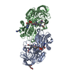
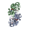
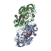
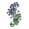
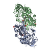
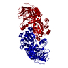
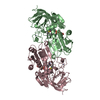
| Title | Ternary complex of Human glutathione-dependent formaldehyde dehydrogenase with S-(hydroxymethyl)glutathione and NADH | ||||||
 Components Components | Alcohol dehydrogenase class III chi chain | ||||||
 Keywords Keywords | OXIDOREDUCTASE / glutathione-dependent formaldehyde dehydrogenase / Class III alcohol dehydrogenase / s-(hydroxymethyl)glutathione | ||||||
| Function / homology |  Function and homology information Function and homology informationformaldehyde dehydrogenase (NAD+) activity / S-nitrosoglutathione reductase (NADH) activity / S-(hydroxymethyl)glutathione dehydrogenase (NADP+) activity / S-(hydroxymethyl)glutathione dehydrogenase (NAD+) activity / S-(hydroxymethyl)glutathione dehydrogenase / fatty acid omega-oxidation / S-(hydroxymethyl)glutathione dehydrogenase [NAD(P)+] activity / response to nitrosative stress / formaldehyde catabolic process / Ethanol oxidation ...formaldehyde dehydrogenase (NAD+) activity / S-nitrosoglutathione reductase (NADH) activity / S-(hydroxymethyl)glutathione dehydrogenase (NADP+) activity / S-(hydroxymethyl)glutathione dehydrogenase (NAD+) activity / S-(hydroxymethyl)glutathione dehydrogenase / fatty acid omega-oxidation / S-(hydroxymethyl)glutathione dehydrogenase [NAD(P)+] activity / response to nitrosative stress / formaldehyde catabolic process / Ethanol oxidation / alcohol dehydrogenase (NAD+) activity / positive regulation of blood pressure / alcohol dehydrogenase / respiratory system process / Oxidoreductases; Acting on the CH-OH group of donors; With NAD+ or NADP+ as acceptor / response to redox state / retinoid metabolic process / fatty acid binding / response to lipopolysaccharide / electron transfer activity / mitochondrion / extracellular exosome / zinc ion binding / identical protein binding / cytosol Similarity search - Function | ||||||
| Biological species |  Homo sapiens (human) Homo sapiens (human) | ||||||
| Method |  X-RAY DIFFRACTION / MOLECULAR REPLACEMENT / Resolution: 2.6 Å X-RAY DIFFRACTION / MOLECULAR REPLACEMENT / Resolution: 2.6 Å | ||||||
 Authors Authors | Sanghani, P.C. / Bosron, W.F. / Hurley, T.D. | ||||||
 Citation Citation | Journal: Biochemistry / Year: 2002 Title: Human glutathione-dependent formaldehyde dehydrogenase. Structural changes associated with Ternary Complex formation Authors: Sanghani, P.C. / Bosron, W.F. / Hurley, T.D. | ||||||
| History |
|
- Structure visualization
| Structure viewer | Molecule: MolmilJmol/JSmol |
|---|
- Downloads & links
-Download
| PDBx/mmCIF format | 1mc5.cif.gz | 161.6 KB | Display | PDBx/mmCIF format |
|---|---|---|---|---|
| PDB format | pdb1mc5.ent.gz | 127.1 KB | Display | PDB format |
| PDBx/mmJSON format | 1mc5.json.gz | Tree view | PDBx/mmJSON format | |
| Others |  Other downloads Other downloads |
-Validation report
| Arichive directory | https://data.pdbj.org/pub/pdb/validation_reports/mc/1mc5ftp://data.pdbj.org/pub/pdb/validation_reports/mc/1mc5 | HTTPS FTP |
|---|
-Related structure data
| Related structure data | 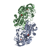 1m6hS S: Starting model for refinement |
|---|---|
| Similar structure data |


-Links
 PDBj
PDBj

- Assembly
| Deposited unit | 
| ||||||||
|---|---|---|---|---|---|---|---|---|---|
| 1 |
| ||||||||
| Unit cell |
|
-Components
-Protein , 1 types, 2 molecules AB
| #1: Protein | Mass: 39772.191 Da / Num. of mol.: 2 Source method: isolated from a genetically manipulated source Source: (gene. exp.) Homo sapiens (human) / Gene: ADH5 / Plasmid: pKK223-3 / Production host:  References: UniProt: P11766, alcohol dehydrogenase, EC: 1.2.1.1 |
|---|
-Non-polymers , 6 types, 406 molecules 


| #2: Chemical | ChemComp-ZN /  Mass: 65.409 Da / Num. of mol.: 4 / Source method: obtained synthetically / Formula: Zn Mass: 65.409 Da / Num. of mol.: 4 / Source method: obtained synthetically / Formula: Zn#3: Chemical | ChemComp-K / |  Mass: 39.098 Da / Num. of mol.: 1 / Source method: obtained synthetically / Formula: K Mass: 39.098 Da / Num. of mol.: 1 / Source method: obtained synthetically / Formula: K#4: Chemical |  Mass: 94.971 Da / Num. of mol.: 3 / Source method: obtained synthetically / Formula: PO4 Mass: 94.971 Da / Num. of mol.: 3 / Source method: obtained synthetically / Formula: PO4#5: Chemical |  Mass: 663.425 Da / Num. of mol.: 2 / Source method: obtained synthetically / Formula: C21H27N7O14P2 / Comment: NAD*YM Mass: 663.425 Da / Num. of mol.: 2 / Source method: obtained synthetically / Formula: C21H27N7O14P2 / Comment: NAD*YM#6: Chemical | ChemComp-AHE / |  Mass: 337.349 Da / Num. of mol.: 1 / Source method: obtained synthetically / Formula: C11H19N3O7S Mass: 337.349 Da / Num. of mol.: 1 / Source method: obtained synthetically / Formula: C11H19N3O7S#7: Water | ChemComp-HOH / | Mass: 18.015 Da / Num. of mol.: 395 / Source method: isolated from a natural source / Formula: H2O |
|---|
-Experimental details
-Experiment
| Experiment | Method: X-RAY DIFFRACTION / Number of used crystals: 1 |
|---|
- Sample preparation
| Crystal | Density Matthews: 3.03 Å3/Da / Density % sol: 59.36 % | ||||||||||||||||||||||||||||||||||||||||||
|---|---|---|---|---|---|---|---|---|---|---|---|---|---|---|---|---|---|---|---|---|---|---|---|---|---|---|---|---|---|---|---|---|---|---|---|---|---|---|---|---|---|---|---|
| Crystal grow | Temperature: 277 K / Method: vapor diffusion, sitting drop / pH: 7.5 Details: PEG8000, Potassium phosphate, zinc sulphate, glutathione, pH 7.5, VAPOR DIFFUSION, SITTING DROP, temperature 277K | ||||||||||||||||||||||||||||||||||||||||||
| Crystal grow | *PLUS Temperature: 4 ℃ / pH: 7.1 | ||||||||||||||||||||||||||||||||||||||||||
| Components of the solutions | *PLUS
|
-Data collection
| Diffraction | Mean temperature: 217 K |
|---|---|
| Diffraction source | Source: ROTATING ANODE / Type: RIGAKU RU200 / Wavelength: 1.5418 Å |
| Detector | Type: RIGAKU RAXIS II / Detector: IMAGE PLATE / Date: Aug 2, 2000 |
| Radiation | Protocol: SINGLE WAVELENGTH / Monochromatic (M) / Laue (L): M / Scattering type: x-ray |
| Radiation wavelength | Wavelength: 1.5418 Å / Relative weight: 1 |
| Reflection | Resolution: 2.6→49 Å / Num. all: 31612 / Num. obs: 27884 / % possible obs: 88.2 % / Observed criterion σ(F): 0 / Observed criterion σ(I): 0 / Redundancy: 4.2 % / Biso Wilson estimate: 53 Å2 / Rmerge(I) obs: 0.035 / Net I/σ(I): 31.2 |
| Reflection shell | Resolution: 2.59→2.69 Å / Redundancy: 1.8 % / Rmerge(I) obs: 0.141 / Mean I/σ(I) obs: 5.9 / Num. unique all: 2155 / % possible all: 70.2 |
| Reflection | *PLUS Lowest resolution: 49 Å / Num. measured all: 117217 |
| Reflection shell | *PLUS % possible obs: 70.2 % |
- Processing
| Software |
| ||||||||||||||||||||||||||||||||||||
|---|---|---|---|---|---|---|---|---|---|---|---|---|---|---|---|---|---|---|---|---|---|---|---|---|---|---|---|---|---|---|---|---|---|---|---|---|---|
| Refinement | Method to determine structure: MOLECULAR REPLACEMENT Starting model: PDB ENTRY 1M6H Resolution: 2.6→48.83 Å / Rfactor Rfree error: 0.007 / Isotropic thermal model: RESTRAINED / Cross valid method: THROUGHOUT / σ(F): 0 / Stereochemistry target values: Engh & Huber
| ||||||||||||||||||||||||||||||||||||
| Solvent computation | Solvent model: FLAT MODEL / Bsol: 42.8432 Å2 / ksol: 0.360354 e/Å3 | ||||||||||||||||||||||||||||||||||||
| Displacement parameters | Biso mean: 35.4 Å2
| ||||||||||||||||||||||||||||||||||||
| Refine analyze |
| ||||||||||||||||||||||||||||||||||||
| Refinement step | Cycle: LAST / Resolution: 2.6→48.83 Å
| ||||||||||||||||||||||||||||||||||||
| Refine LS restraints |
| ||||||||||||||||||||||||||||||||||||
| LS refinement shell | Resolution: 2.6→2.69 Å / Rfactor Rfree error: 0.038 / Total num. of bins used: 10
| ||||||||||||||||||||||||||||||||||||
| Xplor file |
| ||||||||||||||||||||||||||||||||||||
| Refinement | *PLUS Highest resolution: 2.6 Å / Lowest resolution: 49 Å | ||||||||||||||||||||||||||||||||||||
| Solvent computation | *PLUS | ||||||||||||||||||||||||||||||||||||
| Displacement parameters | *PLUS | ||||||||||||||||||||||||||||||||||||
| Refine LS restraints | *PLUS
|
