- PDB-5imk: Nanobody targeting human Vsig4 in Spacegroup C2 -
+
Open data
ID or keywords:
Loading...
-
Basic information
Entry
Database: PDB / ID: 5imk
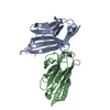
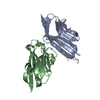
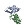
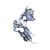
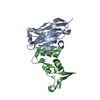
Title
Nanobody targeting human Vsig4 in Spacegroup C2
Components
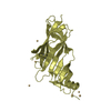
Nanobody
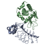
V-set and immunoglobulin domain-containing protein 4
Keywords
IMMUNE SYSTEM / Nanobody / Complement Receptor / Vsig4 CRIg
Function / homology
Function and homology information
negative regulation of complement activation, alternative pathway / negative regulation of macrophage activation / complement component C3b binding / complement activation, alternative pathway / negative regulation of interleukin-2 production / negative regulation of T cell proliferation / protein-containing complex / membrane Similarity search - Function
Mass: 14599.001 Da / Num. of mol.: 1 Source method: isolated from a genetically manipulated source Source: (gene. exp.) Camelidae (mammal) / Production host: Escherichia coli (E. coli)
#2: Protein
V-setandimmunoglobulindomain-containingprotein4 / Protein Z39Ig
Mass: 24606.785 Da / Num. of mol.: 1 / Fragment: UNP residues 19-232 Source method: isolated from a genetically manipulated source Source: (gene. exp.) Homo sapiens (human) / Gene: VSIG4, CRIg, Z39IG, UNQ317/PRO362 / Production host: Escherichia coli (E. coli) / References: UniProt: Q9Y279
In the structure databanks used in Yorodumi, some data are registered as the other names, "COVID-19 virus" and "2019-nCoV". Here are the details of the virus and the list of structure data.
Jan 31, 2019. EMDB accession codes are about to change! (news from PDBe EMDB page)
EMDB accession codes are about to change! (news from PDBe EMDB page)
The allocation of 4 digits for EMDB accession codes will soon come to an end. Whilst these codes will remain in use, new EMDB accession codes will include an additional digit and will expand incrementally as the available range of codes is exhausted. The current 4-digit format prefixed with “EMD-” (i.e. EMD-XXXX) will advance to a 5-digit format (i.e. EMD-XXXXX), and so on. It is currently estimated that the 4-digit codes will be depleted around Spring 2019, at which point the 5-digit format will come into force.
The EM Navigator/Yorodumi systems omit the EMD- prefix.
Related info.:Q: What is EMD? / ID/Accession-code notation in Yorodumi/EM Navigator
Yorodumi is a browser for structure data from EMDB, PDB, SASBDB, etc.
This page is also the successor to EM Navigator detail page, and also detail information page/front-end page for Omokage search.
The word "yorodu" (or yorozu) is an old Japanese word meaning "ten thousand". "mi" (miru) is to see.
Related info.:EMDB / PDB / SASBDB / Comparison of 3 databanks / Yorodumi Search / Aug 31, 2016. New EM Navigator & Yorodumi / Yorodumi Papers / Jmol/JSmol / Function and homology information / Changes in new EM Navigator and Yorodumi
 Movie
Movie Controller


 Open data
 Basic information
 Components
 Keywords
 Function and homology information
 Camelidae (mammal)
 Homo sapiens (human)
 X-RAY DIFFRACTION /
  Authors
 Citation
 Structure visualization
 Downloads & links
 Other downloads


 PDBj
PDBj


 Assembly

 Mass: 18.015 Da / Num. of mol.: 451 / Source method: isolated from a natural source / Formula: H2O
 Sample preparation
 / Beamline: BL19U1 / Wavelength: 0.987 Å
 Processing