Protocol: SINGLE WAVELENGTH / Monochromatic (M) / Laue (L): M / Scattering type: x-ray
Radiation wavelength
Wavelength: 1.0749 Å / Relative weight: 1
Reflection
Resolution: 2.7→49.2 Å / Num. obs: 13917 / % possible obs: 96.8 % / Redundancy: 3.7 % / Net I/σ(I): 23.6
-
Processing
Software
Name
Version
Classification
REFMAC
5.7.0029
refinement
HKL-2000
datareduction
HKL-2000
datascaling
PHENIX
phasing
Refinement
Resolution: 2.7→49.19 Å / Cor.coef. Fo:Fc: 0.924 / Cor.coef. Fo:Fc free: 0.872 / SU B: 14.671 / SU ML: 0.301 / Cross valid method: THROUGHOUT / ESU R: 1.432 / ESU R Free: 0.375 / Details: HYDROGENS HAVE BEEN ADDED IN THE RIDING POSITIONS
Rfactor
Num. reflection
% reflection
Selection details
Rfree
0.27858
743
5.1 %
RANDOM
Rwork
0.22047
-
-
-
obs
0.22342
13917
99.29 %
-
Solvent computation
Ion probe radii: 0.8 Å / Shrinkage radii: 0.8 Å / VDW probe radii: 1.2 Å
 Movie
Movie Controller


 Open data
 Basic information
 Components
 Keywords
 Function and homology information
 X-RAY DIFFRACTION /
  Authors
 United States, 2items
  Citation
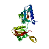
 Structure visualization
 Downloads & links
 Other downloads


 PDBj
PDBj
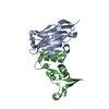
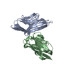
 Assembly


 Sample preparation
 Processing