 Movie
Movie Controller

[English] 日本語
 Yorodumi
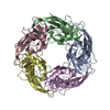
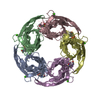
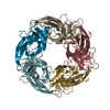
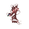
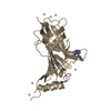
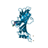
- PDB-3wtn: Crystal Structure of Lymnaea stagnalis Acetylcholine Binding Prot... -
+ Open data

- Basic information
| Entry | Database: PDB / ID: 3wtn | ||||||
|---|---|---|---|---|---|---|---|
| Title | Crystal Structure of Lymnaea stagnalis Acetylcholine Binding Protein Complexed with Desnitro-imidacloprid | ||||||
 Components Components | Acetylcholine-binding protein | ||||||
 Keywords Keywords | SIGNALING PROTEIN / neonicotinoids / nicotinic acetylcholine receptor / imidacloprid / acetylcholine binding | ||||||
| Function / homology |  Function and homology information Function and homology informationsynaptic cleft / extracellular ligand-gated monoatomic ion channel activity / transmembrane signaling receptor activity / membrane Similarity search - Function | ||||||
| Biological species |   Lymnaea stagnalis (great pond snail) Lymnaea stagnalis (great pond snail) | ||||||
| Method |  X-RAY DIFFRACTION / SYNCHROTRON / MOLECULAR REPLACEMENT / Resolution: 2.09 Å X-RAY DIFFRACTION / SYNCHROTRON / MOLECULAR REPLACEMENT / Resolution: 2.09 Å | ||||||
 Authors Authors | Okajima, T. / Ihara, M. / Yamashita, A. / Oda, T. / Matsuda, K. | ||||||
 Citation Citation | Journal: Mol.Pharmacol. / Year: 2014 Title: Studies on an acetylcholine binding protein identify a basic residue in loop G on the beta 1 strand as a new structural determinant of neonicotinoid actions Authors: Ihara, M. / Okajima, T. / Yamashita, A. / Oda, T. / Asano, T. / Matsui, M. / Sattelle, D.B. / Matsuda, K. | ||||||
| History |
|
- Structure visualization
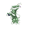
| Structure viewer | Molecule: MolmilJmol/JSmol |
|---|
- Downloads & links
-Download
| PDBx/mmCIF format | 3wtn.cif.gz | 454 KB | Display | PDBx/mmCIF format |
|---|---|---|---|---|
| PDB format | pdb3wtn.ent.gz | 373.2 KB | Display | PDB format |
| PDBx/mmJSON format | 3wtn.json.gz | Tree view | PDBx/mmJSON format | |
| Others |  Other downloads Other downloads |
-Validation report
| Arichive directory | https://data.pdbj.org/pub/pdb/validation_reports/wt/3wtnftp://data.pdbj.org/pub/pdb/validation_reports/wt/3wtn | HTTPS FTP |
|---|
-Related structure data
| Related structure data |  3wthC  3wtiC 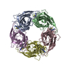 3wtjC  3wtkC  3wtlC  3wtmC  3wtoC  2zjuS S: Starting model for refinement C: citing same article ( |
|---|---|
| Similar structure data |


-Links
 PDBj
PDBj

- Assembly


-Components
| #1: Protein | Mass: 24221.811 Da / Num. of mol.: 10 / Fragment: UNP residues 21-229 Source method: isolated from a genetically manipulated source Source: (gene. exp.) Lymnaea stagnalis (great pond snail) / Plasmid: pPICZ B / Production host:  Pichia pastoris (fungus) / Strain (production host): X-33 / References: UniProt: P58154 Pichia pastoris (fungus) / Strain (production host): X-33 / References: UniProt: P58154#2: Chemical | ChemComp-N2Y / ( 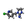  Mass: 210.663 Da / Num. of mol.: 10 / Source method: obtained synthetically / Formula: C9H11ClN4 Mass: 210.663 Da / Num. of mol.: 10 / Source method: obtained synthetically / Formula: C9H11ClN4#3: Chemical | ChemComp-CD /   Mass: 112.411 Da / Num. of mol.: 54 / Source method: obtained synthetically / Formula: Cd Mass: 112.411 Da / Num. of mol.: 54 / Source method: obtained synthetically / Formula: Cd#4: Chemical |   Mass: 22.990 Da / Num. of mol.: 2 / Source method: obtained synthetically / Formula: Na Mass: 22.990 Da / Num. of mol.: 2 / Source method: obtained synthetically / Formula: Na#5: Water | ChemComp-HOH / |  Mass: 18.015 Da / Num. of mol.: 1336 / Source method: isolated from a natural source / Formula: H2O Mass: 18.015 Da / Num. of mol.: 1336 / Source method: isolated from a natural source / Formula: H2OHas protein modification | Y | Sequence details | AUTHORS STATE THAT BEFORE CRYSTALLIZATION, THE N-GLUCOSIDE GROUPS WERE REMOVED BY PEPTIDE-N- ...AUTHORS STATE THAT BEFORE CRYSTALLIZ | |
|---|
-Experimental details
-Experiment
| Experiment | Method: X-RAY DIFFRACTION / Number of used crystals: 1 |
|---|
- Sample preparation
| Crystal | Density Matthews: 2.29 Å3/Da / Density % sol: 46.34 % |
|---|---|
| Crystal grow | Temperature: 293 K / Method: vapor diffusion / pH: 7.5 Details: 1.5M Na acetate, 0.05M CdSO4, 0.1M HEPES-Na (pH 7.5), 0.5mM Desnitro-imidacloprid, VAPOR DIFFUSION, temperature 293K |
-Data collection
| Diffraction | Mean temperature: 100 K |
|---|---|
| Diffraction source | Source: SYNCHROTRON / Site: SPring-8  / Beamline: BL44B2 / Wavelength: 0.9 Å / Beamline: BL44B2 / Wavelength: 0.9 Å |
| Detector | Type: RIGAKU JUPITER 210 / Detector: CCD / Date: Jun 13, 2008 |
| Radiation | Monochromator: Si(111) / Protocol: SINGLE WAVELENGTH / Monochromatic (M) / Laue (L): M / Scattering type: x-ray |
| Radiation wavelength | Wavelength: 0.9 Å / Relative weight: 1 |
| Reflection | Resolution: 2.09→50 Å / Num. all: 137631 / Num. obs: 131277 / % possible obs: 99.2 % / Observed criterion σ(I): -3 / Redundancy: 7.1 % / Biso Wilson estimate: 22.7 Å2 / Rmerge(I) obs: 0.074 |
| Reflection shell | Resolution: 2.09→2.18 Å / Redundancy: 6.4 % / Rmerge(I) obs: 0.463 / Mean I/σ(I) obs: 2.8 / Num. unique all: 12230 / % possible all: 93.7 |
- Processing
| Software |
| ||||||||||||||||||||||||||||||||||||||||||||||||||||||||||||
|---|---|---|---|---|---|---|---|---|---|---|---|---|---|---|---|---|---|---|---|---|---|---|---|---|---|---|---|---|---|---|---|---|---|---|---|---|---|---|---|---|---|---|---|---|---|---|---|---|---|---|---|---|---|---|---|---|---|---|---|---|---|
| Refinement | Method to determine structure: MOLECULAR REPLACEMENT Starting model: 2ZJU Resolution: 2.09→47.84 Å / Rfactor Rfree error: 0.003 / Data cutoff high absF: 2790612.39 / Data cutoff low absF: 0 / Isotropic thermal model: RESTRAINED / Cross valid method: THROUGHOUT / σ(F): 1 / Stereochemistry target values: Engh & Huber / Details: BULK SOLVENT MODEL USED
| ||||||||||||||||||||||||||||||||||||||||||||||||||||||||||||
| Solvent computation | Solvent model: FLAT MODEL / Bsol: 71.3895 Å2 / ksol: 0.4 e/Å3 | ||||||||||||||||||||||||||||||||||||||||||||||||||||||||||||
| Displacement parameters | Biso mean: 38.5 Å2
| ||||||||||||||||||||||||||||||||||||||||||||||||||||||||||||
| Refine analyze |
| ||||||||||||||||||||||||||||||||||||||||||||||||||||||||||||
| Refinement step | Cycle: LAST / Resolution: 2.09→47.84 Å
| ||||||||||||||||||||||||||||||||||||||||||||||||||||||||||||
| Refine LS restraints |
| ||||||||||||||||||||||||||||||||||||||||||||||||||||||||||||
| LS refinement shell | Resolution: 2.09→2.22 Å / Rfactor Rfree error: 0.01 / Total num. of bins used: 6
| ||||||||||||||||||||||||||||||||||||||||||||||||||||||||||||
| Xplor file |
|
