 Movie
Movie Controller

[English] 日本語
 Yorodumi
- PDB-1uw6: X-ray structure of acetylcholine binding protein (AChBP) in compl... -
+ Open data

- Basic information
| Entry | Database: PDB / ID: 1uw6 | ||||||
|---|---|---|---|---|---|---|---|
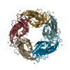
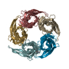
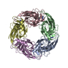
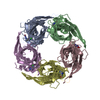
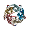
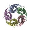
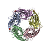
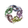
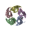
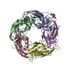
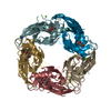
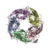
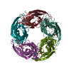
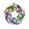
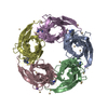
| Title | X-ray structure of acetylcholine binding protein (AChBP) in complex with nicotine | ||||||
 Components Components | ACETYLCHOLINE-BINDING PROTEIN | ||||||
 Keywords Keywords | GLYCOPROTEIN / PENTAMER / IGG FOLD / ACETYLCHOLINE / NICOTINE | ||||||
| Function / homology |  Function and homology information Function and homology informationsynaptic cleft / extracellular ligand-gated monoatomic ion channel activity / transmembrane signaling receptor activity / membrane Similarity search - Function | ||||||
| Biological species |  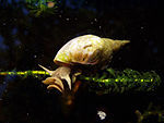 LYMNAEA STAGNALIS (great pond snail) LYMNAEA STAGNALIS (great pond snail) | ||||||
| Method |  X-RAY DIFFRACTION / SYNCHROTRON / MOLECULAR REPLACEMENT / Resolution: 2.2 Å X-RAY DIFFRACTION / SYNCHROTRON / MOLECULAR REPLACEMENT / Resolution: 2.2 Å | ||||||
 Authors Authors | Celie, P.H.N. / Van Rossum-fikkert, S.E. / Van Dijk, W.J. / Brejc, K. / Smit, A.B. / Sixma, T.K. | ||||||
 Citation Citation | Journal: Neuron / Year: 2004 Title: Nicotine and Carbamylcholine Binding to Nicotinic Acetylcholine Receptors as Studied in Achbp Crystal Structures Authors: Celie, P.H.N. / Van Rossum-Fikkert, S.E. / Van Dijk, W.J. / Brejc, K. / Smit, A.B. / Sixma, T.K. #1: Journal: Nature / Year: 2001Title: Crystal Structure of an Ach-Binding Protein Reveals the Ligand-Binding Domain of Nicotinic Receptors Authors: Brejc, K. / Van Dijk, W.J. / Klaassen, R. / Schuurmans, M. / Van Der Oost, J. / Smit, A.B. / Sixma, T.K. #2: Journal: Nature / Year: 2001 Title: A Glia-Derived Acetylcholine-Binding Protein that Modulates Synaptic Transmission Authors: Smit, A.B. / Syed, N.I. / Schaap, D. / Van Minnen, J. / Klumperman, J. / Kits, K.S. / Lodder, H. / Van Der Schors, R.C. / Van Elk, R. / Sorgedrager, B. / Brejc, K. / Sixma, T.K. / Geraerts, W.P.M. | ||||||
| History |
| ||||||
| Remark 700 | SHEET THE SHEET STRUCTURE OF THIS MOLECULE IS BIFURCATED. IN ORDER TO REPRESENT THIS FEATURE IN ... SHEET THE SHEET STRUCTURE OF THIS MOLECULE IS BIFURCATED. IN ORDER TO REPRESENT THIS FEATURE IN THE SHEET RECORDS BELOW, TWO SHEETS ARE DEFINED. |
- Structure visualization
| Structure viewer | Molecule: MolmilJmol/JSmol |
|---|
- Downloads & links
-Download
| PDBx/mmCIF format | 1uw6.cif.gz | 818.9 KB | Display | PDBx/mmCIF format |
|---|---|---|---|---|
| PDB format | pdb1uw6.ent.gz | 687.4 KB | Display | PDB format |
| PDBx/mmJSON format | 1uw6.json.gz | Tree view | PDBx/mmJSON format | |
| Others |  Other downloads Other downloads |
-Validation report
| Arichive directory | https://data.pdbj.org/pub/pdb/validation_reports/uw/1uw6ftp://data.pdbj.org/pub/pdb/validation_reports/uw/1uw6 | HTTPS FTP |
|---|
-Related structure data
| Related structure data |  1uv6C 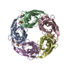 1ux2C 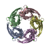 1i9bS S: Starting model for refinement C: citing same article ( |
|---|---|
| Similar structure data |


-Links
 PDBj
PDBj

- Assembly
| Deposited unit | 
| ||||||||||||||||||||||||||||||||||||||||||||||||||||||||||||||||||||||||||||||||
|---|---|---|---|---|---|---|---|---|---|---|---|---|---|---|---|---|---|---|---|---|---|---|---|---|---|---|---|---|---|---|---|---|---|---|---|---|---|---|---|---|---|---|---|---|---|---|---|---|---|---|---|---|---|---|---|---|---|---|---|---|---|---|---|---|---|---|---|---|---|---|---|---|---|---|---|---|---|---|---|---|---|
| 1 | 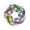
| ||||||||||||||||||||||||||||||||||||||||||||||||||||||||||||||||||||||||||||||||
| 2 | 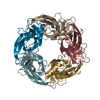
| ||||||||||||||||||||||||||||||||||||||||||||||||||||||||||||||||||||||||||||||||
| 3 | 
| ||||||||||||||||||||||||||||||||||||||||||||||||||||||||||||||||||||||||||||||||
| 4 | 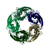
| ||||||||||||||||||||||||||||||||||||||||||||||||||||||||||||||||||||||||||||||||
| Unit cell |
| ||||||||||||||||||||||||||||||||||||||||||||||||||||||||||||||||||||||||||||||||
| Components on special symmetry positions |
| ||||||||||||||||||||||||||||||||||||||||||||||||||||||||||||||||||||||||||||||||
| Noncrystallographic symmetry (NCS) | NCS oper:
|
-Components
| #1: Protein | Mass: 24025.652 Da / Num. of mol.: 20 Source method: isolated from a genetically manipulated source Source: (gene. exp.) LYMNAEA STAGNALIS (great pond snail) / Plasmid: PPIC9 / Production host:  KOMAGATAELLA PASTORIS (fungus) / Strain (production host): GS 115 / References: UniProt: P58154 KOMAGATAELLA PASTORIS (fungus) / Strain (production host): GS 115 / References: UniProt: P58154#2: Chemical | ChemComp-NCT / (   Mass: 162.232 Da / Num. of mol.: 20 / Source method: obtained synthetically / Formula: C10H14N2 / Comment: alkaloid*YM Mass: 162.232 Da / Num. of mol.: 20 / Source method: obtained synthetically / Formula: C10H14N2 / Comment: alkaloid*YM#3: Water | ChemComp-HOH / |  Mass: 18.015 Da / Num. of mol.: 1469 / Source method: isolated from a natural source / Formula: H2O Mass: 18.015 Da / Num. of mol.: 1469 / Source method: isolated from a natural source / Formula: H2OHas protein modification | Y | |
|---|
-Experimental details
-Experiment
| Experiment | Method: X-RAY DIFFRACTION / Number of used crystals: 1 |
|---|
- Sample preparation
| Crystal | Density Matthews: 2.5 Å3/Da / Density % sol: 50 % | ||||||||||||||||||||||||||||||||||||||||||
|---|---|---|---|---|---|---|---|---|---|---|---|---|---|---|---|---|---|---|---|---|---|---|---|---|---|---|---|---|---|---|---|---|---|---|---|---|---|---|---|---|---|---|---|
| Crystal grow | pH: 8 / Details: TRIS PH 8.0, AMMONIUM SULFATE | ||||||||||||||||||||||||||||||||||||||||||
| Crystal grow | *PLUS Method: vapor diffusion, hanging drop | ||||||||||||||||||||||||||||||||||||||||||
| Components of the solutions | *PLUS
|
-Data collection
| Diffraction | Mean temperature: 100 K |
|---|---|
| Diffraction source | Source: SYNCHROTRON / Site: ESRF  / Beamline: ID14-4 / Wavelength: 0.9393 / Beamline: ID14-4 / Wavelength: 0.9393 |
| Detector | Type: ADSC CCD / Detector: CCD / Date: Jun 15, 2002 |
| Radiation | Protocol: SINGLE WAVELENGTH / Monochromatic (M) / Laue (L): M / Scattering type: x-ray |
| Radiation wavelength | Wavelength: 0.9393 Å / Relative weight: 1 |
| Reflection | Resolution: 2.2→50 Å / Num. obs: 236373 / % possible obs: 86 % / Observed criterion σ(I): 1.7 / Redundancy: 2.9 % / Rmerge(I) obs: 0.094 / Net I/σ(I): 9 |
| Reflection shell | Resolution: 2.2→2.28 Å / Redundancy: 1.89 % / Rmerge(I) obs: 0.589 / Mean I/σ(I) obs: 1.7 / % possible all: 38 |
| Reflection | *PLUS Highest resolution: 2.2 Å / Lowest resolution: 50 Å / % possible obs: 86 % / Redundancy: 8 % / Rmerge(I) obs: 0.094 |
| Reflection shell | *PLUS % possible obs: 38 % / Mean I/σ(I) obs: 1.7 |
- Processing
| Software |
| ||||||||||||||||||||
|---|---|---|---|---|---|---|---|---|---|---|---|---|---|---|---|---|---|---|---|---|---|
| Refinement | Method to determine structure: MOLECULAR REPLACEMENT Starting model: PDB ENTRY 1I9B Resolution: 2.2→12 Å / SU B: 8.062 / SU ML: 0.196 / Cross valid method: THROUGHOUT / ESU R: 0.462 / ESU R Free: 0.273
| ||||||||||||||||||||
| Displacement parameters | Biso mean: 21.888 Å2
| ||||||||||||||||||||
| Refinement step | Cycle: LAST / Resolution: 2.2→12 Å
| ||||||||||||||||||||
| Refinement | *PLUS Lowest resolution: 12 Å / Rfactor Rfree: 0.265 / Rfactor Rwork: 0.224 | ||||||||||||||||||||
| Solvent computation | *PLUS | ||||||||||||||||||||
| Displacement parameters | *PLUS | ||||||||||||||||||||
| Refine LS restraints | *PLUS
|
