 Movie
Movie Controller


+ Open data

- Basic information
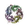
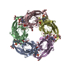
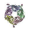
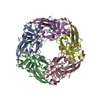
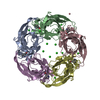
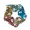
| Entry | Database: PDB / ID: 6sh0 | ||||||
|---|---|---|---|---|---|---|---|
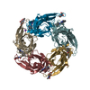
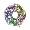
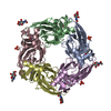
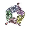
| Title | Crystal structure of AcAChBP in complex with anatoxin | ||||||
 Components Components | Acetylcholine binding protein | ||||||
 Keywords Keywords | SIGNALING PROTEIN / toxin / acetylcholine binding protein | ||||||
| Function / homology |  Function and homology information Function and homology informationextracellular ligand-gated monoatomic ion channel activity / transmembrane signaling receptor activity / membrane / metal ion binding / identical protein binding Similarity search - Function | ||||||
| Biological species |  | ||||||
| Method |  X-RAY DIFFRACTION / MOLECULAR REPLACEMENT / Resolution: 2.5 Å X-RAY DIFFRACTION / MOLECULAR REPLACEMENT / Resolution: 2.5 Å | ||||||
 Authors Authors | Hunter, W.N. / Dawson, A. / Parker, H. | ||||||
 Citation Citation | Journal: Acta Crystallogr.,Sect.F / Year: 2022 Title: Delineating the activity of the potent nicotinic acetylcholine receptor agonists (+)-anatoxin-a and (-)-hosieine-A Authors: Parker, H.P. / Dawson, A. / Jones, M.J. / Yan, R. / Ouyang, J. / Hong, R. / Hunter, W.N. | ||||||
| History |
|
- Structure visualization
| Structure viewer | Molecule: MolmilJmol/JSmol |
|---|
- Downloads & links
-Download
| PDBx/mmCIF format | 6sh0.cif.gz | 433.1 KB | Display | PDBx/mmCIF format |
|---|---|---|---|---|
| PDB format | pdb6sh0.ent.gz | 354.9 KB | Display | PDB format |
| PDBx/mmJSON format | 6sh0.json.gz | Tree view | PDBx/mmJSON format | |
| Others |  Other downloads Other downloads |
-Validation report
| Arichive directory | https://data.pdbj.org/pub/pdb/validation_reports/sh/6sh0ftp://data.pdbj.org/pub/pdb/validation_reports/sh/6sh0 | HTTPS FTP |
|---|
-Related structure data
| Related structure data |  6sgvC  2xysS S: Starting model for refinement C: citing same article ( |
|---|---|
| Similar structure data |


-Links
 PDBj
PDBj

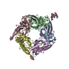
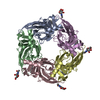
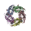
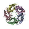
- Assembly
| Deposited unit | 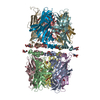
| ||||||||
|---|---|---|---|---|---|---|---|---|---|
| 1 | 
| ||||||||
| 2 | 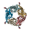
| ||||||||
| Unit cell |
|
-Components
-Protein / Sugars , 2 types, 19 molecules ABCDEFGHIJ
| #1: Protein | Mass: 28326.750 Da / Num. of mol.: 10 Source method: isolated from a genetically manipulated source Source: (gene. exp.) Cell line (production host): Sf9 / Production host:   Spodoptera frugiperda (fall armyworm) / References: UniProt: Q8WSF8 Spodoptera frugiperda (fall armyworm) / References: UniProt: Q8WSF8#2: Sugar | ChemComp-NAG /  Type: D-saccharide, beta linking / Mass: 221.208 Da / Num. of mol.: 9 Type: D-saccharide, beta linking / Mass: 221.208 Da / Num. of mol.: 9Source method: isolated from a genetically manipulated source Formula: C8H15NO6 |
|---|
-Non-polymers , 4 types, 982 molecules 


| #3: Chemical | ChemComp-GOL /  Mass: 92.094 Da / Num. of mol.: 5 / Source method: obtained synthetically / Formula: C3H8O3 Mass: 92.094 Da / Num. of mol.: 5 / Source method: obtained synthetically / Formula: C3H8O3#4: Chemical | ChemComp-4P0 /  Mass: 165.232 Da / Num. of mol.: 10 / Source method: obtained synthetically / Formula: C10H15NO / Feature type: SUBJECT OF INVESTIGATION Mass: 165.232 Da / Num. of mol.: 10 / Source method: obtained synthetically / Formula: C10H15NO / Feature type: SUBJECT OF INVESTIGATION#5: Chemical | ChemComp-ACT /  Mass: 59.044 Da / Num. of mol.: 10 / Source method: obtained synthetically / Formula: C2H3O2 Mass: 59.044 Da / Num. of mol.: 10 / Source method: obtained synthetically / Formula: C2H3O2#6: Water | ChemComp-HOH / | Mass: 18.015 Da / Num. of mol.: 957 / Source method: isolated from a natural source / Formula: H2O |
|---|
-Details
| Has ligand of interest | Y |
|---|---|
| Has protein modification | Y |
-Experimental details
-Experiment
| Experiment | Method: X-RAY DIFFRACTION / Number of used crystals: 1 |
|---|
- Sample preparation
| Crystal | Density Matthews: 3.76 Å3/Da / Density % sol: 67.31 % |
|---|---|
| Crystal grow | Temperature: 298 K / Method: vapor diffusion, hanging drop / pH: 4.6 Details: Reservoir solution: 8% PEG 4K, 0.1 M NaOAc pH 4.6 Protein buffer: 50 mM tris, 250 mM NaCl, pH 7.5, 4mg/ml |
-Data collection
| Diffraction | Mean temperature: 100 K / Serial crystal experiment: N |
|---|---|
| Diffraction source | Source: ROTATING ANODE / Type: RIGAKU MICROMAX-007 HF / Wavelength: 1.5418 Å |
| Detector | Type: RIGAKU SATURN 944+ / Detector: CCD / Date: Dec 15, 2017 |
| Radiation | Monochromator: Si / Protocol: SINGLE WAVELENGTH / Monochromatic (M) / Laue (L): M / Scattering type: x-ray |
| Radiation wavelength | Wavelength: 1.5418 Å / Relative weight: 1 |
| Reflection | Resolution: 2.5→127.87 Å / Num. obs: 119138 / % possible obs: 99.5 % / Redundancy: 11.8 % / Biso Wilson estimate: 23 Å2 / CC1/2: 0.982 / Rmerge(I) obs: 0.228 / Rpim(I) all: 0.089 / Net I/σ(I): 7.4 |
| Reflection shell | Resolution: 2.5→2.54 Å / Redundancy: 9.3 % / Rmerge(I) obs: 0.662 / Mean I/σ(I) obs: 1.8 / Num. unique obs: 5811 / CC1/2: 0.813 / Rpim(I) all: 0.286 / % possible all: 99.3 |
- Processing
| Software |
| ||||||||||||||||||||||||||||||||||||||||||||||||||||||||||||
|---|---|---|---|---|---|---|---|---|---|---|---|---|---|---|---|---|---|---|---|---|---|---|---|---|---|---|---|---|---|---|---|---|---|---|---|---|---|---|---|---|---|---|---|---|---|---|---|---|---|---|---|---|---|---|---|---|---|---|---|---|---|
| Refinement | Method to determine structure: MOLECULAR REPLACEMENT Starting model: 2xys Resolution: 2.5→59.77 Å / Cor.coef. Fo:Fc: 0.893 / Cor.coef. Fo:Fc free: 0.85 / SU B: 11.279 / SU ML: 0.243 / Cross valid method: THROUGHOUT / σ(F): 0 / ESU R: 0.419 / ESU R Free: 0.292 Details: HYDROGENS HAVE BEEN ADDED IN THE RIDING POSITIONS U VALUES : REFINED INDIVIDUALLY
| ||||||||||||||||||||||||||||||||||||||||||||||||||||||||||||
| Solvent computation | Ion probe radii: 0.8 Å / Shrinkage radii: 0.8 Å / VDW probe radii: 1.2 Å | ||||||||||||||||||||||||||||||||||||||||||||||||||||||||||||
| Displacement parameters | Biso max: 72.47 Å2 / Biso mean: 26.137 Å2 / Biso min: 4.96 Å2
| ||||||||||||||||||||||||||||||||||||||||||||||||||||||||||||
| Refinement step | Cycle: final / Resolution: 2.5→59.77 Å
| ||||||||||||||||||||||||||||||||||||||||||||||||||||||||||||
| Refine LS restraints |
| ||||||||||||||||||||||||||||||||||||||||||||||||||||||||||||
| LS refinement shell | Resolution: 2.5→2.565 Å / Rfactor Rfree error: 0 / Total num. of bins used: 20
|
