 Movie
Movie Controller

[English] 日本語
 Yorodumi
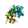
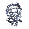
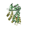
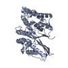
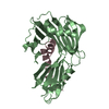
- PDB-6fhe: Highly active enzymes by automated modular backbone assembly and ... -
+ Open data

- Basic information
| Entry | Database: PDB / ID: 6fhe | ||||||
|---|---|---|---|---|---|---|---|
| Title | Highly active enzymes by automated modular backbone assembly and sequence design | ||||||
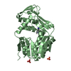
 Components Components | Synthetic construct | ||||||
 Keywords Keywords | Artificial enzyme / Design | ||||||
| Function / homology | Glycosidases / TIM Barrel / Alpha-Beta Barrel / Alpha Beta Function and homology information Function and homology information | ||||||
| Biological species | synthetic construct (others) | ||||||
| Method |  X-RAY DIFFRACTION / MOLECULAR REPLACEMENT / Resolution: 1.93 Å X-RAY DIFFRACTION / MOLECULAR REPLACEMENT / Resolution: 1.93 Å | ||||||
 Authors Authors | Lapidot, G. / Khersonsky, O. / Lipsh, R. / Dym, O. / Albeck, S. / Rogotner, S. / Fleishman, J.S. | ||||||
 Citation Citation | Journal: Nat Commun / Year: 2018 Title: Highly active enzymes by automated combinatorial backbone assembly and sequence design. Authors: Lapidoth, G. / Khersonsky, O. / Lipsh, R. / Dym, O. / Albeck, S. / Rogotner, S. / Fleishman, S.J. | ||||||
| History |
|
- Structure visualization
| Structure viewer | Molecule: MolmilJmol/JSmol |
|---|
- Downloads & links
-Download
| PDBx/mmCIF format | 6fhe.cif.gz | 77.2 KB | Display | PDBx/mmCIF format |
|---|---|---|---|---|
| PDB format | pdb6fhe.ent.gz | 56.9 KB | Display | PDB format |
| PDBx/mmJSON format | 6fhe.json.gz | Tree view | PDBx/mmJSON format | |
| Others |  Other downloads Other downloads |
-Validation report
| Summary document | 6fhe_validation.pdf.gz | 428.8 KB | Display | wwPDB validaton report |
|---|---|---|---|---|
| Full document | 6fhe_full_validation.pdf.gz | 436.2 KB | Display | |
| Data in XML | 6fhe_validation.xml.gz | 13.9 KB | Display | |
| Data in CIF | 6fhe_validation.cif.gz | 18.6 KB | Display | |
| Arichive directory | https://data.pdbj.org/pub/pdb/validation_reports/fh/6fheftp://data.pdbj.org/pub/pdb/validation_reports/fh/6fhe | HTTPS FTP |
-Related structure data
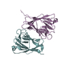
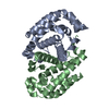
| Related structure data | 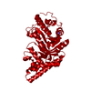 6fhfC 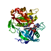 3w2fS S: Starting model for refinement C: citing same article ( |
|---|---|
| Similar structure data |


-Links
 PDBj
PDBj- Assembly
| Deposited unit | 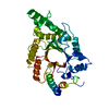
| ||||||||
|---|---|---|---|---|---|---|---|---|---|
| 1 |
| ||||||||
| Unit cell |
|
-Components
| #1: Protein | Mass: 39038.512 Da / Num. of mol.: 1 Source method: isolated from a genetically manipulated source Source: (gene. exp.) synthetic construct (others) / Production host:  |
|---|---|
| #2: Water | ChemComp-HOH /  Mass: 18.015 Da / Num. of mol.: 71 / Source method: isolated from a natural source / Formula: H2O Mass: 18.015 Da / Num. of mol.: 71 / Source method: isolated from a natural source / Formula: H2O |
-Experimental details
-Experiment
| Experiment | Method: X-RAY DIFFRACTION / Number of used crystals: 1 |
|---|
- Sample preparation
| Crystal | Density Matthews: 2.47 Å3/Da / Density % sol: 50.21 % |
|---|---|
| Crystal grow | Temperature: 292 K / Method: vapor diffusion, hanging drop / Details: 8% PEG 3,350 0.05M Tri-sodium cidrate pH=5.8 |
-Data collection
| Diffraction | Mean temperature: 100 K |
|---|---|
| Diffraction source | Source: ROTATING ANODE / Type: RIGAKU RUH3R / Wavelength: 1.541 Å |
| Detector | Type: RIGAKU / Detector: AREA DETECTOR / Date: Jul 12, 2017 |
| Radiation | Protocol: SINGLE WAVELENGTH / Monochromatic (M) / Laue (L): M / Scattering type: x-ray |
| Radiation wavelength | Wavelength: 1.541 Å / Relative weight: 1 |
| Reflection | Resolution: 1.93→51.02 Å / Num. obs: 30737 / % possible obs: 99 % / Observed criterion σ(F): 0 / Redundancy: 6.4 % / Biso Wilson estimate: 37.6 Å2 / Rsym value: 0.047 / Net I/σ(I): 7.6 |
| Reflection shell | Resolution: 1.93→1.97 Å / Redundancy: 6.3 % / Mean I/σ(I) obs: 1 / Num. unique obs: 2057 / Rsym value: 0.66 / % possible all: 99.9 |
- Processing
| Software |
| ||||||||||||||||
|---|---|---|---|---|---|---|---|---|---|---|---|---|---|---|---|---|---|
| Refinement | Method to determine structure: MOLECULAR REPLACEMENT Starting model: 3W2F Resolution: 1.93→42.02 Å / Cross valid method: FREE R-VALUE
| ||||||||||||||||
| Displacement parameters | Biso mean: 44.26 Å2 | ||||||||||||||||
| Refinement step | Cycle: LAST / Resolution: 1.93→42.02 Å
|
