 Movie
Movie Controller


+ Open data

- Basic information
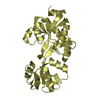
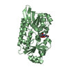
| Entry | Database: PDB / ID: 6l9w | |||||||||
|---|---|---|---|---|---|---|---|---|---|---|
| Title | Crystal structure of mouse TIFA (T9E/C36S mutant) | |||||||||
 Components Components | TRAF-interacting protein with FHA domain-containing protein A | |||||||||
 Keywords Keywords | SIGNALING PROTEIN / FHA domain | |||||||||
| Function / homology |  Function and homology information Function and homology informationAlpha-protein kinase 1 signaling pathway / TAK1-dependent IKK and NF-kappa-B activation / cytoplasmic pattern recognition receptor signaling pathway / tumor necrosis factor-mediated signaling pathway / protein homooligomerization / positive regulation of canonical NF-kappaB signal transduction / innate immune response / cytoplasm Similarity search - Function | |||||||||
| Biological species |  | |||||||||
| Method |  X-RAY DIFFRACTION / SYNCHROTRON / MOLECULAR REPLACEMENT / Resolution: 2.9 Å X-RAY DIFFRACTION / SYNCHROTRON / MOLECULAR REPLACEMENT / Resolution: 2.9 Å | |||||||||
 Authors Authors | Nakamura, T. / Yamagata, Y. | |||||||||
| Funding support |  Japan, 2items Japan, 2items
| |||||||||
 Citation Citation | Journal: Sci Rep / Year: 2020 Title: Structural analysis of TIFA: Insight into TIFA-dependent signal transduction in innate immunity. Authors: Nakamura, T. / Hashikawa, C. / Okabe, K. / Yokote, Y. / Chirifu, M. / Toma-Fukai, S. / Nakamura, N. / Matsuo, M. / Kamikariya, M. / Okamoto, Y. / Gohda, J. / Akiyama, T. / Semba, K. / ...Authors: Nakamura, T. / Hashikawa, C. / Okabe, K. / Yokote, Y. / Chirifu, M. / Toma-Fukai, S. / Nakamura, N. / Matsuo, M. / Kamikariya, M. / Okamoto, Y. / Gohda, J. / Akiyama, T. / Semba, K. / Ikemizu, S. / Otsuka, M. / Inoue, J.I. / Yamagata, Y. #1: Journal: To Be PublishedTitle: Crystallization and preliminary X-ray analysis of mouse TIFA Authors: Nakamura, T. / Inoue, J. / Yamagata, Y. | |||||||||
| History |
|
- Structure visualization
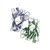
| Structure viewer | Molecule: MolmilJmol/JSmol |
|---|
- Downloads & links
-Download
| PDBx/mmCIF format | 6l9w.cif.gz | 182 KB | Display | PDBx/mmCIF format |
|---|---|---|---|---|
| PDB format | pdb6l9w.ent.gz | 144.3 KB | Display | PDB format |
| PDBx/mmJSON format | 6l9w.json.gz | Tree view | PDBx/mmJSON format | |
| Others |  Other downloads Other downloads |
-Validation report
| Arichive directory | https://data.pdbj.org/pub/pdb/validation_reports/l9/6l9wftp://data.pdbj.org/pub/pdb/validation_reports/l9/6l9w | HTTPS FTP |
|---|
-Related structure data
| Related structure data | 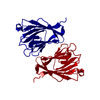 6l9uSC  6l9vC S: Starting model for refinement C: citing same article ( |
|---|---|
| Similar structure data |


-Links
 PDBj
PDBj

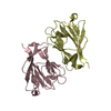
- Assembly
| Deposited unit | 
| ||||||||
|---|---|---|---|---|---|---|---|---|---|
| 1 | 
| ||||||||
| 2 | 
| ||||||||
| 3 | 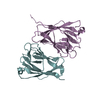
| ||||||||
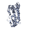
| Unit cell |
|
-Components
| #1: Protein | Mass: 22663.414 Da / Num. of mol.: 6 / Mutation: T9E,C36S Source method: isolated from a genetically manipulated source Source: (gene. exp.)  |
|---|
-Experimental details
-Experiment
| Experiment | Method: X-RAY DIFFRACTION / Number of used crystals: 1 |
|---|
- Sample preparation
| Crystal | Density Matthews: 2.46 Å3/Da / Density % sol: 50.1 % |
|---|---|
| Crystal grow | Temperature: 293 K / Method: vapor diffusion, hanging drop / pH: 6.4 / Details: MES pH 6.4, MPD |
-Data collection
| Diffraction | Mean temperature: 100 K / Serial crystal experiment: N |
|---|---|
| Diffraction source | Source: SYNCHROTRON / Site: SPring-8 / Beamline: BL41XU / Wavelength: 1 Å |
| Detector | Type: RAYONIX MX225HE / Detector: CCD / Date: Jan 30, 2013 |
| Radiation | Protocol: SINGLE WAVELENGTH / Monochromatic (M) / Laue (L): M / Scattering type: x-ray |
| Radiation wavelength | Wavelength: 1 Å / Relative weight: 1 |
| Reflection | Resolution: 2.9→37.29 Å / Num. obs: 28175 / % possible obs: 95.7 % / Redundancy: 3.7 % / Rmerge(I) obs: 0.084 / Net I/σ(I): 23.3 |
| Reflection shell | Resolution: 2.9→3 Å / Rmerge(I) obs: 0.294 / Num. unique obs: 2170 |
- Processing
| Software |
| ||||||||||||||||||||||||||||||||||||||||||||||||||||||||||||||||||
|---|---|---|---|---|---|---|---|---|---|---|---|---|---|---|---|---|---|---|---|---|---|---|---|---|---|---|---|---|---|---|---|---|---|---|---|---|---|---|---|---|---|---|---|---|---|---|---|---|---|---|---|---|---|---|---|---|---|---|---|---|---|---|---|---|---|---|---|
| Refinement | Method to determine structure: MOLECULAR REPLACEMENT Starting model: 6L9U Resolution: 2.9→37.29 Å / SU ML: 0.4 / Cross valid method: THROUGHOUT / σ(F): 1.35 / Phase error: 32.79
| ||||||||||||||||||||||||||||||||||||||||||||||||||||||||||||||||||
| Solvent computation | Shrinkage radii: 0.9 Å / VDW probe radii: 1.11 Å | ||||||||||||||||||||||||||||||||||||||||||||||||||||||||||||||||||
| Displacement parameters | Biso max: 137.62 Å2 / Biso mean: 61.7854 Å2 / Biso min: 17.11 Å2 | ||||||||||||||||||||||||||||||||||||||||||||||||||||||||||||||||||
| Refinement step | Cycle: final / Resolution: 2.9→37.29 Å
| ||||||||||||||||||||||||||||||||||||||||||||||||||||||||||||||||||
| LS refinement shell | Refine-ID: X-RAY DIFFRACTION / Rfactor Rfree error: 0
|
