 Movie
Movie Controller

[English] 日本語
 Yorodumi
- PDB-5nny: Crystal structure of the phosphatase domain from the Legionella e... -
+ Open data

- Basic information
| Entry | Database: PDB / ID: 5nny | ||||||
|---|---|---|---|---|---|---|---|
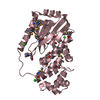
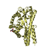
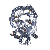
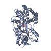
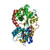
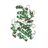
| Title | Crystal structure of the phosphatase domain from the Legionella effector WipB | ||||||
 Components Components | WipB | ||||||
 Keywords Keywords | HYDROLASE / Legionella effector / Serine/Threonine phosphatase / phosphoesterase fold | ||||||
| Function / homology | : / WipA-like, phosphatase domain / Domain of unknown function DUF5617 / Domain of unknown function (DUF5617) / phosphatase activity / WipB Function and homology information Function and homology information | ||||||
| Biological species |   Legionella pneumophila (bacteria) Legionella pneumophila (bacteria) | ||||||
| Method |  X-RAY DIFFRACTION / SYNCHROTRON / MOLECULAR REPLACEMENT / Resolution: 1.7 Å X-RAY DIFFRACTION / SYNCHROTRON / MOLECULAR REPLACEMENT / Resolution: 1.7 Å | ||||||
 Authors Authors | Pinotsis, N. / Waksman, G. / Prevost, M.S. | ||||||
| Funding support |  Belgium, 1items Belgium, 1items
| ||||||
 Citation Citation | Journal: Sci Rep / Year: 2017 Title: The Legionella effector WipB is a translocated Ser/Thr phosphatase that targets the host lysosomal nutrient sensing machinery. Authors: Prevost, M.S. / Pinotsis, N. / Dumoux, M. / Hayward, R.D. / Waksman, G. #1: Journal: To Be PublishedTitle: Structure of WipA reveals a novel tyrosine protein phosphatase effector from Legionella pneumophila Authors: Pinotsis, N. / Waksman, G. / Prevost, M.S. | ||||||
| History |
|
- Structure visualization
| Structure viewer | Molecule: MolmilJmol/JSmol |
|---|
- Downloads & links
-Download
| PDBx/mmCIF format | 5nny.cif.gz | 265.3 KB | Display | PDBx/mmCIF format |
|---|---|---|---|---|
| PDB format | pdb5nny.ent.gz | 213.8 KB | Display | PDB format |
| PDBx/mmJSON format | 5nny.json.gz | Tree view | PDBx/mmJSON format | |
| Others |  Other downloads Other downloads |
-Validation report
| Arichive directory | https://data.pdbj.org/pub/pdb/validation_reports/nn/5nnyftp://data.pdbj.org/pub/pdb/validation_reports/nn/5nny | HTTPS FTP |
|---|
-Related structure data
| Related structure data |  5n6xS S: Starting model for refinement |
|---|---|
| Similar structure data |


-Links
 PDBj
PDBj- Assembly
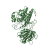
| Deposited unit | 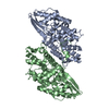
| ||||||||
|---|---|---|---|---|---|---|---|---|---|
| 1 | 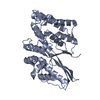
| ||||||||
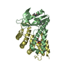
| 2 | 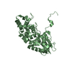
| ||||||||
| Unit cell |
|
-Components
| #1: Protein | Mass: 36337.312 Da / Num. of mol.: 2 / Fragment: Phosphatase domain, UNP Residues 25-344 Source method: isolated from a genetically manipulated source Source: (gene. exp.) Legionella pneumophila (bacteria) / Gene: wipB / Production host: #2: Water | ChemComp-HOH / |  Mass: 18.015 Da / Num. of mol.: 484 / Source method: isolated from a natural source / Formula: H2O Mass: 18.015 Da / Num. of mol.: 484 / Source method: isolated from a natural source / Formula: H2O |
|---|
-Experimental details
-Experiment
| Experiment | Method: X-RAY DIFFRACTION / Number of used crystals: 1 |
|---|
- Sample preparation
| Crystal | Density Matthews: 2.12 Å3/Da / Density % sol: 41.93 % |
|---|---|
| Crystal grow | Temperature: 289 K / Method: vapor diffusion / pH: 7.5 / Details: 0.23 M ammonium citrate dibasic and 22% PEG3000 |
-Data collection
| Diffraction | Mean temperature: 100 K |
|---|---|
| Diffraction source | Source: SYNCHROTRON / Site: PETRA III, EMBL c/o DESY  / Beamline: P13 (MX1) / Wavelength: 0.99999 Å / Beamline: P13 (MX1) / Wavelength: 0.99999 Å |
| Detector | Type: DECTRIS PILATUS3 S 6M / Detector: PIXEL / Date: Nov 26, 2016 |
| Radiation | Protocol: SINGLE WAVELENGTH / Monochromatic (M) / Laue (L): M / Scattering type: x-ray |
| Radiation wavelength | Wavelength: 0.99999 Å / Relative weight: 1 |
| Reflection | Resolution: 1.7→48.832 Å / Num. obs: 66193 / % possible obs: 99.3 % / Redundancy: 3.6 % / CC1/2: 0.996 / Rmerge(I) obs: 0.116 / Net I/σ(I): 8.73 |
| Reflection shell | Resolution: 1.7→1.74 Å / Mean I/σ(I) obs: 1.21 / Num. unique obs: 4732 / CC1/2: 0.48 / % possible all: 97.1 |
- Processing
| Software |
| ||||||||||||||||||||||||||||||||||||||||||||||||||||||||||||||||||||||||||||||||||||||||||||||||||||||||||||||||||||||||||||||||||||||||||||||||||||||||||||||||||||||||
|---|---|---|---|---|---|---|---|---|---|---|---|---|---|---|---|---|---|---|---|---|---|---|---|---|---|---|---|---|---|---|---|---|---|---|---|---|---|---|---|---|---|---|---|---|---|---|---|---|---|---|---|---|---|---|---|---|---|---|---|---|---|---|---|---|---|---|---|---|---|---|---|---|---|---|---|---|---|---|---|---|---|---|---|---|---|---|---|---|---|---|---|---|---|---|---|---|---|---|---|---|---|---|---|---|---|---|---|---|---|---|---|---|---|---|---|---|---|---|---|---|---|---|---|---|---|---|---|---|---|---|---|---|---|---|---|---|---|---|---|---|---|---|---|---|---|---|---|---|---|---|---|---|---|---|---|---|---|---|---|---|---|---|---|---|---|---|---|---|---|
| Refinement | Method to determine structure: MOLECULAR REPLACEMENT Starting model: 5N6X Resolution: 1.7→48.832 Å / SU ML: 0.24 / Cross valid method: FREE R-VALUE / σ(F): 1.36 / Phase error: 23.94 / Stereochemistry target values: ML
| ||||||||||||||||||||||||||||||||||||||||||||||||||||||||||||||||||||||||||||||||||||||||||||||||||||||||||||||||||||||||||||||||||||||||||||||||||||||||||||||||||||||||
| Solvent computation | Shrinkage radii: 0.9 Å / VDW probe radii: 1.11 Å / Solvent model: FLAT BULK SOLVENT MODEL | ||||||||||||||||||||||||||||||||||||||||||||||||||||||||||||||||||||||||||||||||||||||||||||||||||||||||||||||||||||||||||||||||||||||||||||||||||||||||||||||||||||||||
| Refinement step | Cycle: LAST / Resolution: 1.7→48.832 Å
| ||||||||||||||||||||||||||||||||||||||||||||||||||||||||||||||||||||||||||||||||||||||||||||||||||||||||||||||||||||||||||||||||||||||||||||||||||||||||||||||||||||||||
| Refine LS restraints |
| ||||||||||||||||||||||||||||||||||||||||||||||||||||||||||||||||||||||||||||||||||||||||||||||||||||||||||||||||||||||||||||||||||||||||||||||||||||||||||||||||||||||||
| LS refinement shell |
| ||||||||||||||||||||||||||||||||||||||||||||||||||||||||||||||||||||||||||||||||||||||||||||||||||||||||||||||||||||||||||||||||||||||||||||||||||||||||||||||||||||||||
| Refinement TLS params. | S33: -0 Å ° / Method: refined / Refine-ID: X-RAY DIFFRACTION
| ||||||||||||||||||||||||||||||||||||||||||||||||||||||||||||||||||||||||||||||||||||||||||||||||||||||||||||||||||||||||||||||||||||||||||||||||||||||||||||||||||||||||
| Refinement TLS group |
|
