| Entry | Database: PDB / ID: 6ff0
|
|---|
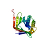
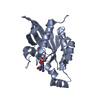
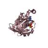
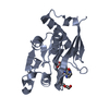
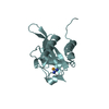
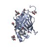
| Title | Ryegrass mottle virus serine protease domain fused with VPg domain |
|---|
 Components Components | Serine protease domain fused with VPg domain |
|---|
 Keywords Keywords | VIRAL PROTEIN / serine protease / viral protease / viral polypeptide / viral genome-linked protein |
|---|
| Function / homology |  Function and homology information Function and homology information
host cell membrane / serine-type endopeptidase activity / viral translational frameshifting / viral RNA genome replication / nucleotide binding / RNA-directed RNA polymerase activity / DNA-templated transcription / proteolysis / RNA binding / membraneSimilarity search - Function Peptidase S39B, luteovirus / Peptidase family S39 domain profile. / RNA-directed RNA polymerase, luteovirus / Viral RNA-directed RNA-polymerase / RNA-directed RNA polymerase, catalytic domain / RdRp of positive ssRNA viruses catalytic domain profile. / Peptidase S1, PA clan, chymotrypsin-like fold / Peptidase S1, PA clan / DNA/RNA polymerase superfamilySimilarity search - Domain/homology |
|---|
| Biological species |  Ryegrass mottle virus Ryegrass mottle virus |
|---|
| Method |  X-RAY DIFFRACTION / SYNCHROTRON / MOLECULAR REPLACEMENT / Resolution: 2.1 Å X-RAY DIFFRACTION / SYNCHROTRON / MOLECULAR REPLACEMENT / Resolution: 2.1 Å |
|---|
 Authors Authors | Kalnins, G. |
|---|
 Citation Citation | Journal: Int J Mol Sci / Year: 2023
Title: VPg Impact on Ryegrass Mottle Virus Serine-like 3C Protease Proteolysis and Structure.
Authors: Kalnins, G. / Ludviga, R. / Kalnciema, I. / Resevica, G. / Zeltina, V. / Bogans, J. / Tars, K. / Zeltins, A. / Balke, I. |
|---|
| History | | Deposition | Jan 3, 2018 | Deposition site: PDBE / Processing site: PDBE |
|---|
| Revision 1.0 | Apr 17, 2019 | Provider: repository / Type: Initial release |
|---|
| Revision 1.1 | Apr 5, 2023 | Group: Database references / Category: citation / citation_author / database_2
Item: _citation.country / _citation.journal_abbrev ..._citation.country / _citation.journal_abbrev / _citation.journal_id_CSD / _citation.journal_id_ISSN / _citation.journal_volume / _citation.pdbx_database_id_DOI / _citation.pdbx_database_id_PubMed / _citation.title / _citation.year / _database_2.pdbx_DOI / _database_2.pdbx_database_accession |
|---|
| Revision 1.2 | Feb 7, 2024 | Group: Data collection / Refinement description
Category: chem_comp_atom / chem_comp_bond / pdbx_initial_refinement_model |
|---|
| Revision 1.3 | Oct 23, 2024 | Group: Structure summary / Category: pdbx_entry_details / pdbx_modification_feature |
|---|
|
|---|
 Movie
Movie Controller

 Yorodumi
 Open data
 Basic information
 Structure visualization
 Downloads & links
 Other downloads
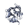


 PDBj
PDBj Assembly
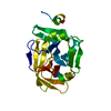

 Mass: 18.015 Da / Num. of mol.: 51 / Source method: isolated from a natural source / Formula: H2O
 Sample preparation
 / Beamline: 14.1 / Wavelength: 0.96411 Å
 Processing