[English] 日本語
Yorodumi
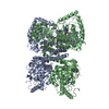
- PDB-6fe8: Cryo-EM structure of the core Centromere Binding Factor 3 complex -

+
Open data


ID or keywords:

Loading...

-
Basic information

Entry
Database: PDB / ID: 6fe8
TitleCryo-EM structure of the core Centromere Binding Factor 3 complex
Components
  • Centromere DNA-binding protein complex CBF3 subunit B
  • Centromere DNA-binding protein complex CBF3 subunit C
  • Suppressor of kinetochore protein 1
KeywordsDNA BINDING PROTEIN / Centromere / CDEIII-binding / LRR domain
Function / homology
Function and homology information


RAVE complex / Iron uptake and transport / CBF3 complex / regulation of transcription by galactose / regulation of sulfur amino acid metabolic process / cellular response to methylmercury / vacuolar proton-transporting V-type ATPase complex assembly / septin ring assembly / centromeric DNA binding / regulation of exit from mitosis ...RAVE complex / Iron uptake and transport / CBF3 complex / regulation of transcription by galactose / regulation of sulfur amino acid metabolic process / cellular response to methylmercury / vacuolar proton-transporting V-type ATPase complex assembly / septin ring assembly / centromeric DNA binding / regulation of exit from mitosis / kinetochore assembly / vacuolar acidification / positive regulation of D-glucose transmembrane transport / protein neddylation / regulation of metabolic process / mitochondrial fusion / mitotic intra-S DNA damage checkpoint signaling / silent mating-type cassette heterochromatin formation / DNA binding, bending / exit from mitosis / SCF ubiquitin ligase complex / SCF-dependent proteasomal ubiquitin-dependent protein catabolic process / FBXL7 down-regulates AURKA during mitotic entry and in early mitosis / mitotic spindle assembly checkpoint signaling / Orc1 removal from chromatin / DNA replication origin binding / cullin family protein binding / Antigen processing: Ubiquitination & Proteasome degradation / regulation of protein-containing complex assembly / subtelomeric heterochromatin formation / endomembrane system / negative regulation of cytoplasmic translation / regulation of mitotic cell cycle / kinetochore / G1/S transition of mitotic cell cycle / G2/M transition of mitotic cell cycle / mitotic cell cycle / ubiquitin-dependent protein catabolic process / protein-containing complex assembly / chromosome, telomeric region / DNA-binding transcription factor activity, RNA polymerase II-specific / protein ubiquitination / zinc ion binding / identical protein binding / nucleus / cytoplasm
Similarity search - Function
Centromere DNA-binding protein complex CBF3 subunit B, C-terminal / : / Centromere DNA-binding protein complex CBF3 subunit B / Zn(2)-C6 fungal-type DNA-binding domain signature. / Fungal Zn(2)-Cys(6) binuclear cluster domain / Zn(2)-C6 fungal-type DNA-binding domain superfamily / Zn(2)-C6 fungal-type DNA-binding domain profile. / GAL4-like Zn(II)2Cys6 (or C6 zinc) binuclear cluster DNA-binding domain / Zn(2)-C6 fungal-type DNA-binding domain / SKP1 component, dimerisation ...Centromere DNA-binding protein complex CBF3 subunit B, C-terminal / : / Centromere DNA-binding protein complex CBF3 subunit B / Zn(2)-C6 fungal-type DNA-binding domain signature. / Fungal Zn(2)-Cys(6) binuclear cluster domain / Zn(2)-C6 fungal-type DNA-binding domain superfamily / Zn(2)-C6 fungal-type DNA-binding domain profile. / GAL4-like Zn(II)2Cys6 (or C6 zinc) binuclear cluster DNA-binding domain / Zn(2)-C6 fungal-type DNA-binding domain / SKP1 component, dimerisation / S-phase kinase-associated protein 1 / SKP1-like, dimerisation domain superfamily / Skp1 family, dimerisation domain / Potassium Channel Kv1.1; Chain A / Potassium Channel Kv1.1; Chain A / S-phase kinase-associated protein 1-like / SKP1 component, POZ domain / Skp1 family, tetramerisation domain / Found in Skp1 protein family / SKP1/BTB/POZ domain superfamily / Leucine-rich repeat domain superfamily / 2-Layer Sandwich / Alpha Beta
Similarity search - Domain/homology
Centromere DNA-binding protein complex CBF3 subunit C / Centromere DNA-binding protein complex CBF3 subunit B / Suppressor of kinetochore protein 1
Similarity search - Component
Biological speciesSaccharomyces cerevisiae (brewer's yeast)
MethodELECTRON MICROSCOPY / single particle reconstruction / cryo EM / Resolution: 4.1 Å
AuthorsZhang, W.J. / Lukoynova, N. / Miah, S. / Vaughan, C.K.
Funding support United Kingdom, 1items
OrganizationGrant numberCountry
Biotechnology and Biological Sciences Research CouncilBB/J007595/1 United Kingdom
CitationJournal: Cell Rep / Year: 2018
Title: Insights into Centromere DNA Bending Revealed by the Cryo-EM Structure of the Core Centromere Binding Factor 3 with Ndc10.
Authors: Wenjuan Zhang / Natalya Lukoyanova / Shomon Miah / Jonathan Lucas / Cara K Vaughan /
Abstract: The centromere binding factor 3 (CBF3) complex binds the third centromere DNA element in organisms with point centromeres, such as S. cerevisiae. It is an essential complex for assembly of the ...The centromere binding factor 3 (CBF3) complex binds the third centromere DNA element in organisms with point centromeres, such as S. cerevisiae. It is an essential complex for assembly of the kinetochore in these organisms, as it facilitates genetic centromere specification and allows association of all other kinetochore components. We determined high-resolution structures of the core complex of CBF3 alone and in association with a monomeric construct of Ndc10, using cryoelectron microscopy (cryo-EM). We identify the DNA-binding site of the complex and present a model in which CBF3 induces a tight bend in centromeric DNA, thus facilitating assembly of the centromeric nucleosome.
History
DepositionDec 30, 2017Deposition site: PDBE / Processing site: PDBE
Revision 1.0Aug 1, 2018Provider: repository / Type: Initial release
Revision 1.1Oct 3, 2018Group: Data collection / Refinement description / Category: refine
Revision 1.2Dec 11, 2019Group: Other / Category: atom_sites
Item: _atom_sites.fract_transf_matrix[1][1] / _atom_sites.fract_transf_matrix[2][2] / _atom_sites.fract_transf_matrix[3][3]

-
Structure visualization

Movie
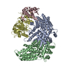
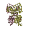
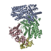
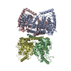
  • Deposited structure unit
  • Imaged by Jmol
  • Download
  • Superimposition on EM map
  • EMDB-4241
  • Imaged by UCSF Chimera
  • Download
Movie viewer
Structure viewerMolecule:
MolmilJmol/JSmol

Downloads & links

-
Assembly

Deposited unit
A: Centromere DNA-binding protein complex CBF3 subunit B
B: Centromere DNA-binding protein complex CBF3 subunit B
C: Suppressor of kinetochore protein 1
D: Centromere DNA-binding protein complex CBF3 subunit C


Theoretical massNumber of molelcules
Total (without water)220,3674
Polymers220,3674
Non-polymers00
Water00
1


  • Idetical with deposited unit
  • defined by author&software
  • Evidence: gel filtration, It shows a single and symmetric peak at an elution volume corresponding to 220kDa.
TypeNameSymmetry operationNumber
identity operation1_555x,y,z1
Buried area13630 Å2
ΔGint-88 kcal/mol
Surface area66160 Å2
MethodPISA

-
Components

#1: Protein Centromere DNA-binding protein complex CBF3 subunit B / Centromere protein 3


Mass: 68454.125 Da / Num. of mol.: 2
Source method: isolated from a genetically manipulated source
Details: Model is numbered according to
Source: (gene. exp.) Saccharomyces cerevisiae (brewer's yeast)
Gene: CEP3, CBF3, CBF3B, CSL1, YMR168C, YM8520.17C / Production host: Saccharomyces cerevisiae (brewer's yeast) / References: UniProt: P40969
#2: Protein Suppressor of kinetochore protein 1 / Centromere DNA-binding protein complex CBF3 subunit D / E3 ubiquitin ligase complex SCF subunit SKP1


Mass: 22558.451 Da / Num. of mol.: 1
Source method: isolated from a genetically manipulated source
Source: (gene. exp.) Saccharomyces cerevisiae (brewer's yeast)
Gene: SKP1, CBF3D, YDR328C, D9798.14 / Production host: Saccharomyces cerevisiae (brewer's yeast) / References: UniProt: P52286
#3: Protein Centromere DNA-binding protein complex CBF3 subunit C / Chromosome transmission fidelity protein 13 / Kinetochore protein CTF13


Mass: 60899.961 Da / Num. of mol.: 1
Source method: isolated from a genetically manipulated source
Source: (gene. exp.) Saccharomyces cerevisiae (brewer's yeast)
Gene: CTF13, CBF3C, YMR094W, YM6543.01, YM9582.19 / Production host: Saccharomyces cerevisiae (brewer's yeast) / References: UniProt: P35203

-
Experimental details

-
Experiment

ExperimentMethod: ELECTRON MICROSCOPY
EM experimentAggregation state: PARTICLE / 3D reconstruction method: single particle reconstruction

-
Sample preparation

ComponentName: The core Centromere Binding Factor 3 complex / Type: COMPLEX
Details: The core CBF3 complex, recombinantly expressed in Saccharomyces cerevisiae. It comprises a Cep3 homodimer, in which the binuclear zinc cluster domains are truncated, and full length Skp1 and Ctf13 components.
Entity ID: all / Source: RECOMBINANT
Molecular weightValue: 0.22 MDa / Experimental value: NO
Source (natural)Organism: Saccharomyces cerevisiae (brewer's yeast)
Source (recombinant)Organism: Saccharomyces cerevisiae (brewer's yeast)
Buffer solutionpH: 8
Buffer component
IDConc.NameFormulaBuffer-ID
1200 mMsodium chlorideNaCl1
215 mMTris base1
32 mMDTT1
SpecimenConc.: 0.12 mg/ml / Embedding applied: NO / Shadowing applied: NO / Staining applied: NO / Vitrification applied: YES
Details: The sample is homogeneous and well-dispersed on grids.
Specimen supportGrid material: GOLD / Grid mesh size: 300 divisions/in. / Grid type: Quantifoil R1.2/1.3
VitrificationInstrument: FEI VITROBOT MARK I / Cryogen name: ETHANE / Humidity: 100 % / Chamber temperature: 277 K

-
Electron microscopy imaging

Experimental equipment
Model: Titan Krios / Image courtesy: FEI Company
MicroscopyModel: FEI TITAN KRIOS
Electron gunElectron source: FIELD EMISSION GUN / Accelerating voltage: 300 kV / Illumination mode: FLOOD BEAM
Electron lensMode: BRIGHT FIELD / Calibrated magnification: 47170 X / Cs: 2.7 mm
Specimen holderCryogen: NITROGEN / Specimen holder model: FEI TITAN KRIOS AUTOGRID HOLDER
Image recording

Imaging-ID: 1 / Detector mode: COUNTING / Film or detector model: GATAN K2 SUMMIT (4k x 4k) / Num. of grids imaged: 1

IDAverage exposure time (sec.)Electron dose (e/Å2)Num. of real images
10.44.61236
20.3754.061101
Image scansMovie frames/image: 40 / Used frames/image: 1-40

-
Processing

SoftwareName: PHENIX / Version: 1.13_2992: / Classification: refinement
EM software
IDNameVersionCategoryDetailsImage processing-ID
1Gautomatchparticle selectionGautomatch was used to pick a subset of the initial dataset for making references and for further automatic particle picking of the initial dataset1
3EPU1.9.1image acquisitionData collection
5CTFFIND4CTF correctionCTF parameters were estimated using CTFFIND41
8UCSF Chimera1.8.1model fittingUsed for initial placement of Cep3 and BTB/POZ domain of Skp1
11Coot0.8.8model fittingUsed for ab initio building of all of Ctf13, the C-terminal section of Skp1, and parts of Cep3 where new density was visible or where it deviated from the crystal structure coordinates
12PHENIX1.13model refinementReal Space Refinement Protocol
13REFMACmodel refinement
14RELION2initial Euler assignment1
16RELION2final Euler assignment1
18RELION2classification1
20RELION23D reconstruction1
22Gautomatchparticle selectionGautomatch was used to pick a subset of the initial dataset for making references and for further automatic particle picking of the initial dataset2
33CTFFIND4CTF correctionCTF parameters were estimated using CTFFIND42
35RELION2initial Euler assignment2
37RELION2final Euler assignment2
39RELION2classification2
41RELION23D reconstruction2
Image processing
IDImage recording-IDDetails
11The selected images were high-pass filtered and normalized.
22The selected images were high-pass filtered and normalized.
CTF correction
IDEM image processing-IDType
11PHASE FLIPPING AND AMPLITUDE CORRECTION
22PHASE FLIPPING AND AMPLITUDE CORRECTION
Particle selection
IDImage processing-IDNum. of particles selected
11139303
22289281
Symmetry
IDImage processing-IDEntry-IDPoint symmetry
116FE8C1 (asymmetric)
226FE8C1 (asymmetric)
3D reconstruction

Algorithm: FOURIER SPACE / Entry-ID: 6FE8 / Resolution method: FSC 0.143 CUT-OFF / Symmetry type: POINT

IDResolution (Å)Num. of particlesImage processing-IDDetailsNum. of class averages
14.16939213D auto-refinement of all the good particles from 2D classification, provided a reconstruction at 4.7 angstrom overall resolution. After post-process by RELION and sharpened by a negative B-factor using an automated procedure resulting in a 4.1 angstrom reconstruction.4
23.71876062187, 606 particles were in the best two 3D classes. 3D auto-refinement of the 3D joined classes against the corresponding particles resulted in a final reconstruction.2
Atomic model buildingProtocol: AB INITIO MODEL / Space: REAL / Target criteria: Cross-correlation coefficient
Atomic model building
IDPDB-IDPdb chain-ID 3D fitting-IDPdb chain residue range
12VEQA148-608
21NEXA1
RefinementResolution: 3.7→127.2 Å / Cor.coef. Fo:Fc: 0.884 / ESU R: 1.068
Stereochemistry target values: MAXIMUM LIKELIHOOD WITH PHASES
Details: HYDROGENS HAVE BEEN ADDED IN THE RIDING POSITIONS
RfactorNum. reflection% reflection
Rwork0.29358 --
obs0.29358 62402 100 %
Solvent computationIon probe radii: 0.8 Å / Shrinkage radii: 0.8 Å / VDW probe radii: 1.2 Å / Solvent model: MASK
Displacement parametersBiso mean: 117.316 Å2
Baniso -1Baniso -2Baniso -3
1-0.92 Å21.76 Å2-5.08 Å2
2---2.74 Å2-1.5 Å2
3---1.82 Å2
Refinement stepCycle: 1 / Total: 12393
Refine LS restraints
Refine-IDTypeDev idealDev ideal targetNumber
ELECTRON MICROSCOPYr_bond_refined_d0.020.01912682
ELECTRON MICROSCOPYr_bond_other_d00.0211685
ELECTRON MICROSCOPYr_angle_refined_deg2.251.95517218
ELECTRON MICROSCOPYr_angle_other_deg3.765326900
ELECTRON MICROSCOPYr_dihedral_angle_1_deg7.60551535
ELECTRON MICROSCOPYr_dihedral_angle_2_deg30.62523.657566
ELECTRON MICROSCOPYr_dihedral_angle_3_deg15.126152140
ELECTRON MICROSCOPYr_dihedral_angle_4_deg15.5631568
ELECTRON MICROSCOPYr_chiral_restr0.1490.21966
ELECTRON MICROSCOPYr_gen_planes_refined0.0090.0213935
ELECTRON MICROSCOPYr_gen_planes_other0.0130.022754
ELECTRON MICROSCOPYr_nbd_refined
ELECTRON MICROSCOPYr_nbd_other
ELECTRON MICROSCOPYr_nbtor_refined
ELECTRON MICROSCOPYr_nbtor_other
ELECTRON MICROSCOPYr_xyhbond_nbd_refined
ELECTRON MICROSCOPYr_xyhbond_nbd_other
ELECTRON MICROSCOPYr_metal_ion_refined
ELECTRON MICROSCOPYr_metal_ion_other
ELECTRON MICROSCOPYr_symmetry_vdw_refined
ELECTRON MICROSCOPYr_symmetry_vdw_other
ELECTRON MICROSCOPYr_symmetry_hbond_refined
ELECTRON MICROSCOPYr_symmetry_hbond_other
ELECTRON MICROSCOPYr_symmetry_metal_ion_refined
ELECTRON MICROSCOPYr_symmetry_metal_ion_other
ELECTRON MICROSCOPYr_mcbond_it18.68910.9986176
ELECTRON MICROSCOPYr_mcbond_other18.68310.9976175
ELECTRON MICROSCOPYr_mcangle_it30.38616.3937699
ELECTRON MICROSCOPYr_mcangle_other30.38416.3957700
ELECTRON MICROSCOPYr_scbond_it26.28913.1336506
ELECTRON MICROSCOPYr_scbond_other26.28713.1336506
ELECTRON MICROSCOPYr_scangle_it
ELECTRON MICROSCOPYr_scangle_other41.65818.7279520
ELECTRON MICROSCOPYr_long_range_B_refined50.88614953
ELECTRON MICROSCOPYr_long_range_B_other50.84614949
ELECTRON MICROSCOPYr_rigid_bond_restr
ELECTRON MICROSCOPYr_sphericity_free
ELECTRON MICROSCOPYr_sphericity_bonded
LS refinement shellResolution: 3.7→3.796 Å / Total num. of bins used: 20
RfactorNum. reflection% reflection
Rwork0.615 4633 -
Rfree-0 -
obs--100 %

+
About Yorodumi

-
News

-
Feb 9, 2022. New format data for meta-information of EMDB entries

New format data for meta-information of EMDB entries

  • Version 3 of the EMDB header file is now the official format.
  • The previous official version 1.9 will be removed from the archive.

Related info.:EMDB header

External links:wwPDB to switch to version 3 of the EMDB data model

-
Aug 12, 2020. Covid-19 info

Covid-19 info

URL: https://pdbj.org/emnavi/covid19.php

New page: Covid-19 featured information page in EM Navigator.

Related info.:Covid-19 info / Mar 5, 2020. Novel coronavirus structure data

+
Mar 5, 2020. Novel coronavirus structure data

Novel coronavirus structure data

Related info.:Yorodumi Speices / Aug 12, 2020. Covid-19 info

External links:COVID-19 featured content - PDBj / Molecule of the Month (242):Coronavirus Proteases

+
Jan 31, 2019. EMDB accession codes are about to change! (news from PDBe EMDB page)

EMDB accession codes are about to change! (news from PDBe EMDB page)

  • The allocation of 4 digits for EMDB accession codes will soon come to an end. Whilst these codes will remain in use, new EMDB accession codes will include an additional digit and will expand incrementally as the available range of codes is exhausted. The current 4-digit format prefixed with “EMD-” (i.e. EMD-XXXX) will advance to a 5-digit format (i.e. EMD-XXXXX), and so on. It is currently estimated that the 4-digit codes will be depleted around Spring 2019, at which point the 5-digit format will come into force.
  • The EM Navigator/Yorodumi systems omit the EMD- prefix.

Related info.:Q: What is EMD? / ID/Accession-code notation in Yorodumi/EM Navigator

External links:EMDB Accession Codes are Changing Soon! / Contact to PDBj

+
Jul 12, 2017. Major update of PDB

Major update of PDB

  • wwPDB released updated PDB data conforming to the new PDBx/mmCIF dictionary.
  • This is a major update changing the version number from 4 to 5, and with Remediation, in which all the entries are updated.
  • In this update, many items about electron microscopy experimental information are reorganized (e.g. em_software).
  • Now, EM Navigator and Yorodumi are based on the updated data.

External links:wwPDB Remediation / Enriched Model Files Conforming to OneDep Data Standards Now Available in the PDB FTP Archive

-
Yorodumi

Thousand views of thousand structures

  • Yorodumi is a browser for structure data from EMDB, PDB, SASBDB, etc.
  • This page is also the successor to EM Navigator detail page, and also detail information page/front-end page for Omokage search.
  • The word "yorodu" (or yorozu) is an old Japanese word meaning "ten thousand". "mi" (miru) is to see.

Related info.:EMDB / PDB / SASBDB / Comparison of 3 databanks / Yorodumi Search / Aug 31, 2016. New EM Navigator & Yorodumi / Yorodumi Papers / Jmol/JSmol / Function and homology information / Changes in new EM Navigator and Yorodumi

Read more