 Movie
Movie Controller


+ Open data

- Basic information
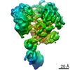
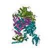
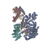
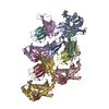
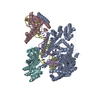
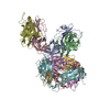
| Entry | Database: PDB / ID: 6eny | ||||||||||||
|---|---|---|---|---|---|---|---|---|---|---|---|---|---|
| Title | Structure of the human PLC editing module | ||||||||||||
 Components Components |
| ||||||||||||
 Keywords Keywords | IMMUNE SYSTEM / adaptive immunity / antigen processing / chaperone / MHC class I | ||||||||||||
| Function / homology |  Function and homology information Function and homology informationMHC class Ib protein complex assembly / peptide antigen stabilization / Tapasin-ERp57 complex / response to biphenyl / Calnexin/calreticulin cycle / MHC class I protein complex binding / cytolytic granule / protein disulfide-isomerase / antigen processing and presentation of exogenous peptide antigen via MHC class I, TAP-dependent / nuclear receptor-mediated glucocorticoid signaling pathway ...MHC class Ib protein complex assembly / peptide antigen stabilization / Tapasin-ERp57 complex / response to biphenyl / Calnexin/calreticulin cycle / MHC class I protein complex binding / cytolytic granule / protein disulfide-isomerase / antigen processing and presentation of exogenous peptide antigen via MHC class I, TAP-dependent / nuclear receptor-mediated glucocorticoid signaling pathway / Assembly of Viral Components at the Budding Site / positive regulation of dendritic cell chemotaxis / ATF6 (ATF6-alpha) activates chaperone genes / negative regulation of trophoblast cell migration / cortical granule / regulation of meiotic nuclear division / negative regulation of retinoic acid receptor signaling pathway / cellular response to electrical stimulus / : / complement component C1q complex binding / endoplasmic reticulum quality control compartment / protein folding in endoplasmic reticulum / sarcoplasmic reticulum lumen / disulfide oxidoreductase activity / response to peptide / negative regulation of intracellular steroid hormone receptor signaling pathway / TAP2 binding / TAP1 binding / regulation of protein complex stability / nuclear export signal receptor activity / C-type glycerophospholipase activity / retrograde vesicle-mediated transport, Golgi to endoplasmic reticulum / cardiac muscle cell differentiation / cortical actin cytoskeleton organization / cellular response to interleukin-7 / positive regulation of memory T cell activation / T cell mediated cytotoxicity directed against tumor cell target / response to glycoside / positive regulation of extrinsic apoptotic signaling pathway / positive regulation of CD8-positive, alpha-beta T cell activation / CD8-positive, alpha-beta T cell activation / positive regulation of CD8-positive, alpha-beta T cell proliferation / Scavenging by Class A Receptors / Scavenging by Class F Receptors / nuclear androgen receptor binding / cellular response to lithium ion / antigen processing and presentation of endogenous peptide antigen via MHC class I via ER pathway, TAP-dependent / negative regulation of neuron differentiation / TAP complex binding / antigen processing and presentation of exogenous peptide antigen via MHC class I / protein disulfide isomerase activity / Golgi medial cisterna / response to testosterone / lncRNA binding / CD8 receptor binding / smooth endoplasmic reticulum / hormone binding / protection from natural killer cell mediated cytotoxicity / beta-2-microglobulin binding / protein-disulfide reductase activity / endoplasmic reticulum exit site / TAP binding / MHC class I protein binding / molecular sequestering activity / detection of bacterium / antigen processing and presentation of endogenous peptide antigen via MHC class Ib / antigen processing and presentation of endogenous peptide antigen via MHC class I via ER pathway, TAP-independent / protein localization to nucleus / T cell receptor binding / extrinsic apoptotic signaling pathway / phagocytic vesicle / ERAD pathway / peptide binding / endocytic vesicle lumen / positive regulation of cell cycle / positive regulation of substrate adhesion-dependent cell spreading / endoplasmic reticulum-Golgi intermediate compartment membrane / protein export from nucleus / protein folding chaperone / positive regulation of endothelial cell migration / positive regulation of phagocytosis / early endosome lumen / response to endoplasmic reticulum stress / Nef mediated downregulation of MHC class I complex cell surface expression / DAP12 interactions / acrosomal vesicle / Endosomal/Vacuolar pathway / T cell mediated cytotoxicity / Antigen Presentation: Folding, assembly and peptide loading of class I MHC / lumenal side of endoplasmic reticulum membrane / regulation of iron ion transport / cellular response to iron(III) ion / negative regulation of iron ion transport / negative regulation of forebrain neuron differentiation / antigen processing and presentation of exogenous protein antigen via MHC class Ib, TAP-dependent / peptide antigen assembly with MHC class I protein complex / ER to Golgi transport vesicle membrane / regulation of erythrocyte differentiation / response to molecule of bacterial origin / HFE-transferrin receptor complex Similarity search - Function | ||||||||||||
| Biological species |  Homo sapiens (human) Homo sapiens (human) | ||||||||||||
| Method | ELECTRON MICROSCOPY / single particle reconstruction / cryo EM / Resolution: 5.8 Å | ||||||||||||
 Authors Authors | Trowitzsch, S. / Januliene, D. / Blees, A. / Moeller, A. / Tampe, R. | ||||||||||||
| Funding support |  Germany, 2items Germany, 2items
| ||||||||||||
 Citation Citation | Journal: Nature / Year: 2017 Title: Structure of the human MHC-I peptide-loading complex. Authors: Andreas Blees / Dovile Januliene / Tommy Hofmann / Nicole Koller / Carla Schmidt / Simon Trowitzsch / Arne Moeller / Robert Tampé / Abstract: The peptide-loading complex (PLC) is a transient, multisubunit membrane complex in the endoplasmic reticulum that is essential for establishing a hierarchical immune response. The PLC coordinates ...The peptide-loading complex (PLC) is a transient, multisubunit membrane complex in the endoplasmic reticulum that is essential for establishing a hierarchical immune response. The PLC coordinates peptide translocation into the endoplasmic reticulum with loading and editing of major histocompatibility complex class I (MHC-I) molecules. After final proofreading in the PLC, stable peptide-MHC-I complexes are released to the cell surface to evoke a T-cell response against infected or malignant cells. Sampling of different MHC-I allomorphs requires the precise coordination of seven different subunits in a single macromolecular assembly, including the transporter associated with antigen processing (TAP1 and TAP2, jointly referred to as TAP), the oxidoreductase ERp57, the MHC-I heterodimer, and the chaperones tapasin and calreticulin. The molecular organization of and mechanistic events that take place in the PLC are unknown owing to the heterogeneous composition and intrinsically dynamic nature of the complex. Here, we isolate human PLC from Burkitt's lymphoma cells using an engineered viral inhibitor as bait and determine the structure of native PLC by electron cryo-microscopy. Two endoplasmic reticulum-resident editing modules composed of tapasin, calreticulin, ERp57, and MHC-I are centred around TAP in a pseudo-symmetric orientation. A multivalent chaperone network within and across the editing modules establishes the proofreading function at two lateral binding platforms for MHC-I molecules. The lectin-like domain of calreticulin senses the MHC-I glycan, whereas the P domain reaches over the MHC-I peptide-binding pocket towards ERp57. This arrangement allows tapasin to facilitate peptide editing by clamping MHC-I. The translocation pathway of TAP opens out into a large endoplasmic reticulum lumenal cavity, confined by the membrane entry points of tapasin and MHC-I. Two lateral windows channel the antigenic peptides to MHC-I. Structures of PLC captured at distinct assembly states provide mechanistic insight into the recruitment and release of MHC-I. Our work defines the molecular symbiosis of an ABC transporter and an endoplasmic reticulum chaperone network in MHC-I assembly and provides insight into the onset of the adaptive immune response. | ||||||||||||
| History |
|
- Structure visualization
| Movie |
Movie viewer |
|---|---|
| Structure viewer | Molecule: MolmilJmol/JSmol |
- Downloads & links
-Download
| PDBx/mmCIF format | 6eny.cif.gz | 257.1 KB | Display | PDBx/mmCIF format |
|---|---|---|---|---|
| PDB format | pdb6eny.ent.gz | 168 KB | Display | PDB format |
| PDBx/mmJSON format | 6eny.json.gz | Tree view | PDBx/mmJSON format | |
| Others |  Other downloads Other downloads |
-Validation report
| Arichive directory | https://data.pdbj.org/pub/pdb/validation_reports/en/6enyftp://data.pdbj.org/pub/pdb/validation_reports/en/6eny | HTTPS FTP |
|---|
-Related structure data
| Related structure data | 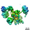 3906MC 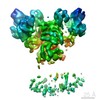 3904C 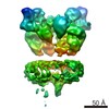 3905C M: map data used to model this data C: citing same article ( |
|---|---|
| Similar structure data |


-Links
 PDBj
PDBj


- Assembly
| Deposited unit | 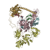
|
|---|---|
| 1 |
|
-Components
-Protein , 5 types, 5 molecules BCDFG
| #1: Protein | Mass: 11748.160 Da / Num. of mol.: 1 / Source method: isolated from a natural source / Source: (natural) Homo sapiens (human) / References: UniProt: P61769 |
|---|---|
| #2: Protein | Mass: 45761.184 Da / Num. of mol.: 1 / Source method: isolated from a natural source / Source: (natural) Homo sapiens (human) / References: UniProt: O15533 |
| #3: Protein | Mass: 54341.102 Da / Num. of mol.: 1 / Source method: isolated from a natural source / Source: (natural) Homo sapiens (human) / References: UniProt: P30101, protein disulfide-isomerase |
| #4: Protein | Mass: 38363.535 Da / Num. of mol.: 1 / Source method: isolated from a natural source / Source: (natural) Homo sapiens (human) / References: UniProt: P04439 |
| #5: Protein | Mass: 46507.145 Da / Num. of mol.: 1 / Source method: isolated from a natural source / Source: (natural) Homo sapiens (human) / References: UniProt: P27797 |
-Sugars , 2 types, 2 molecules
| #6: Polysaccharide | 2-acetamido-2-deoxy-beta-D-glucopyranose-(1-4)-2-acetamido-2-deoxy-beta-D-glucopyranose Source method: isolated from a genetically manipulated source |
|---|---|
| #7: Polysaccharide | beta-D-glucopyranose-(1-3)-alpha-D-mannopyranose-(1-2)-alpha-D-mannopyranose-(1-2)-alpha-D-mannopyranose Type: oligosaccharide / Mass: 666.578 Da / Num. of mol.: 1 Source method: isolated from a genetically manipulated source |
-Experimental details
-Experiment
| Experiment | Method: ELECTRON MICROSCOPY |
|---|---|
| EM experiment | Aggregation state: PARTICLE / 3D reconstruction method: single particle reconstruction |
- Sample preparation
| Component | Name: Protein Complex / Type: COMPLEX / Entity ID: #1-#5 / Source: NATURAL |
|---|---|
| Molecular weight | Experimental value: NO |
| Source (natural) | Organism: Homo sapiens (human) |
| Buffer solution | pH: 7.5 |
| Specimen | Conc.: 2 mg/ml / Embedding applied: NO / Shadowing applied: NO / Staining applied: NO / Vitrification applied: YES |
| Specimen support | Grid type: C-flat-2/2 |
| Vitrification | Instrument: FEI VITROBOT MARK IV / Cryogen name: ETHANE / Humidity: 100 % / Chamber temperature: 277 K |
- Electron microscopy imaging
| Experimental equipment |  Model: Titan Krios / Image courtesy: FEI Company |
|---|---|
| Microscopy | Model: FEI TITAN KRIOS |
| Electron gun | Electron source:  FIELD EMISSION GUN / Accelerating voltage: 300 kV / Illumination mode: FLOOD BEAM FIELD EMISSION GUN / Accelerating voltage: 300 kV / Illumination mode: FLOOD BEAM |
| Electron lens | Mode: BRIGHT FIELD |
| Image recording | Electron dose: 55 e/Å2 / Detector mode: COUNTING / Film or detector model: GATAN K2 QUANTUM (4k x 4k) |
- Processing
| EM software |
| ||||||||||||||||||||||||||||||||||||
|---|---|---|---|---|---|---|---|---|---|---|---|---|---|---|---|---|---|---|---|---|---|---|---|---|---|---|---|---|---|---|---|---|---|---|---|---|---|
| CTF correction | Details: CTF correction was performed internally in Relion and Frealign Type: NONE | ||||||||||||||||||||||||||||||||||||
| Symmetry | Point symmetry: C1 (asymmetric) | ||||||||||||||||||||||||||||||||||||
| 3D reconstruction | Resolution: 5.8 Å / Resolution method: FSC 0.143 CUT-OFF / Num. of particles: 141078 / Symmetry type: POINT | ||||||||||||||||||||||||||||||||||||
| Refinement | Highest resolution: 5.8 Å |
