 Movie
Movie Controller

[English] 日本語
 Yorodumi
- PDB-6cx1: Cryo-EM structure of Seneca Valley Virus-Anthrax Toxin Receptor 1... -
+ Open data

- Basic information
| Entry | Database: PDB / ID: 6cx1 | ||||||
|---|---|---|---|---|---|---|---|
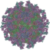
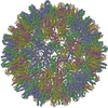
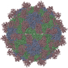
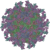
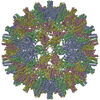
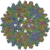
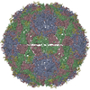
| Title | Cryo-EM structure of Seneca Valley Virus-Anthrax Toxin Receptor 1 complex | ||||||
 Components Components |
| ||||||
 Keywords Keywords | VIRUS / Virus-receptor complex / Picornavirus / Senecavirus / Anthrax Toxin Receptor | ||||||
| Function / homology |  Function and homology information Function and homology informationsymbiont-mediated suppression of host cytoplasmic pattern recognition receptor signaling pathway via inhibition of IRF7 activity / host cell nucleolus / adhesion receptor-mediated virion attachment to host cell / filopodium membrane / negative regulation of extracellular matrix assembly / symbiont-mediated suppression of host TRAF-mediated signal transduction / blood vessel development / lamellipodium membrane / symbiont-mediated suppression of host cytoplasmic pattern recognition receptor signaling pathway via inhibition of RIG-I activity / Uptake and function of anthrax toxins ...symbiont-mediated suppression of host cytoplasmic pattern recognition receptor signaling pathway via inhibition of IRF7 activity / host cell nucleolus / adhesion receptor-mediated virion attachment to host cell / filopodium membrane / negative regulation of extracellular matrix assembly / symbiont-mediated suppression of host TRAF-mediated signal transduction / blood vessel development / lamellipodium membrane / symbiont-mediated suppression of host cytoplasmic pattern recognition receptor signaling pathway via inhibition of RIG-I activity / Uptake and function of anthrax toxins / positive regulation of epidermal growth factor receptor signaling pathway / collagen binding / substrate adhesion-dependent cell spreading / symbiont-mediated suppression of host cytoplasmic pattern recognition receptor signaling pathway via inhibition of MAVS activity / picornain 3C / T=pseudo3 icosahedral viral capsid / host cell cytoplasmic vesicle membrane / actin filament binding / transmembrane signaling receptor activity / signaling receptor activity / channel activity / actin cytoskeleton organization / symbiont-mediated suppression of host cytoplasmic pattern recognition receptor signaling pathway via inhibition of TBK1 activity / monoatomic ion transmembrane transport / symbiont-mediated suppression of host toll-like receptor signaling pathway / symbiont-mediated suppression of host cytoplasmic pattern recognition receptor signaling pathway via inhibition of IRF3 activity / entry receptor-mediated virion attachment to host cell / cysteine-type deubiquitinase activity / ubiquitinyl hydrolase 1 / RNA helicase activity / endosome membrane / RNA helicase / external side of plasma membrane / RNA-directed RNA polymerase / cysteine-type endopeptidase activity / viral RNA genome replication / RNA-directed RNA polymerase activity / symbiont entry into host cell / virion attachment to host cell / structural molecule activity / cell surface / DNA-templated transcription / ATP hydrolysis activity / proteolysis / RNA binding / ATP binding / metal ion binding / plasma membrane Similarity search - Function | ||||||
| Biological species |  Homo sapiens (human) Homo sapiens (human) Senecavirus A Senecavirus A | ||||||
| Method | ELECTRON MICROSCOPY / single particle reconstruction / cryo EM / Resolution: 3.8 Å | ||||||
 Authors Authors | Jayawardena, N. / Burga, L. / Easingwood, R. / Takizawa, Y. / Wolf, M. / Bostina, M. | ||||||
 Citation Citation | Journal: Proc Natl Acad Sci U S A / Year: 2018 Title: Structural basis for anthrax toxin receptor 1 recognition by Seneca Valley Virus. Authors: Nadishka Jayawardena / Laura N Burga / Richard A Easingwood / Yoshimasa Takizawa / Matthias Wolf / Mihnea Bostina /   Abstract: Recently, the use of oncolytic viruses in cancer therapy has become a realistic therapeutic option. Seneca Valley Virus (SVV) is a newly discovered picornavirus, which has earned a significant ...Recently, the use of oncolytic viruses in cancer therapy has become a realistic therapeutic option. Seneca Valley Virus (SVV) is a newly discovered picornavirus, which has earned a significant reputation as a potent oncolytic agent. Anthrax toxin receptor 1 (ANTXR1), one of the cellular receptors for the protective antigen secreted by , has been identified as the high-affinity cellular receptor for SVV. Here, we report the structure of the SVV-ANTXR1 complex determined by single-particle cryo-electron microscopy analysis at near-atomic resolution. This is an example of a shared receptor structure between a mammalian virus and a bacterial toxin. Our structure shows that ANTXR1 decorates the outer surface of the SVV capsid and interacts with the surface-exposed BC loop and loop II of VP1, "the puff" of VP2 and "the knob" of VP3. Comparison of the receptor-bound capsid structure with the native capsid structure reveals that receptor binding induces minor conformational changes in SVV capsid structure, suggesting the role of ANTXR1 as an attachment receptor. Furthermore, our results demonstrate that the capsid footprint on the receptor is not conserved in anthrax toxin receptor 2 (ANTXR2), thereby providing a molecular mechanism for explaining the exquisite selectivity of SVV for ANTXR1. | ||||||
| History |
|
- Structure visualization
| Movie |
Movie viewer |
|---|---|
| Structure viewer | Molecule: MolmilJmol/JSmol |
- Downloads & links
-Download
| PDBx/mmCIF format | 6cx1.cif.gz | 188 KB | Display | PDBx/mmCIF format |
|---|---|---|---|---|
| PDB format | pdb6cx1.ent.gz | 146.3 KB | Display | PDB format |
| PDBx/mmJSON format | 6cx1.json.gz | Tree view | PDBx/mmJSON format | |
| Others |  Other downloads Other downloads |
-Validation report
| Arichive directory | https://data.pdbj.org/pub/pdb/validation_reports/cx/6cx1ftp://data.pdbj.org/pub/pdb/validation_reports/cx/6cx1 | HTTPS FTP |
|---|
-Related structure data
| Related structure data |  7772MC M: map data used to model this data C: citing same article ( |
|---|---|
| Similar structure data |


-Links
 PDBj
PDBj


- Assembly
| Deposited unit | 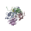
|
|---|---|
| 1 | x 60
|
| 2 |
|
| 3 | x 5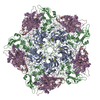
|
| 4 | x 6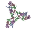
|
| 5 | 
|
| Symmetry | Point symmetry: (Schoenflies symbol: I (icosahedral)) |
-Components
| #1: Protein | Mass: 21026.668 Da / Num. of mol.: 1 Source method: isolated from a genetically manipulated source Source: (gene. exp.) Homo sapiens (human) / Gene: ANTXR1, ATR, TEM8 / Production host: Homo sapiens (human) / References: UniProt: Q9H6X2 |
|---|---|
| #2: Protein | Mass: 28459.969 Da / Num. of mol.: 1 / Fragment: residues 674-931 / Source method: isolated from a natural source / Source: (natural) Senecavirus A / References: UniProt: A0A1U9IRU2, UniProt: Q155Z9*PLUS |
| #3: Protein | Mass: 29870.316 Da / Num. of mol.: 1 / Fragment: residues 162-429 / Source method: isolated from a natural source / Source: (natural) Senecavirus A / References: UniProt: A0A1U9IRU2, UniProt: Q155Z9*PLUS |
| #4: Protein | Mass: 26353.938 Da / Num. of mol.: 1 / Fragment: residues 435-672 / Source method: isolated from a natural source / Source: (natural) Senecavirus A / References: UniProt: A0A1U9IRU2, UniProt: Q155Z9*PLUS |
| #5: Protein | Mass: 5998.467 Da / Num. of mol.: 1 / Fragment: residues 93-150 / Source method: isolated from a natural source / Source: (natural) Senecavirus A / References: UniProt: A0A218L148, UniProt: Q155Z9*PLUS |
-Experimental details
-Experiment
| Experiment | Method: ELECTRON MICROSCOPY |
|---|---|
| EM experiment | Aggregation state: PARTICLE / 3D reconstruction method: single particle reconstruction |
- Sample preparation
| Component | Name: Seneca Valley Virus-Anthrax Toxin Receptor 1 complex / Type: VIRUS / Entity ID: all / Source: MULTIPLE SOURCES |
|---|---|
| Molecular weight | Value: 6.5 MDa / Experimental value: NO |
| Source (natural) | Organism: Senecavirus A |
| Details of virus | Empty: NO / Enveloped: NO / Isolate: STRAIN / Type: VIRION |
| Virus shell | Name: Capsid / Diameter: 300 nm / Triangulation number (T number): 3 |
| Buffer solution | pH: 7.4 |
| Buffer component | Name: Phosphate Buffer |
| Specimen | Conc.: 0.2 mg/ml / Embedding applied: NO / Shadowing applied: NO / Staining applied: NO / Vitrification applied: YES / Details: A homogenous sample containing full capsids |
| Specimen support | Grid mesh size: 300 divisions/in. / Grid type: Quantifoil R1.2/1.3 |
| Vitrification | Instrument: FEI VITROBOT MARK IV / Cryogen name: ETHANE / Humidity: 100 % / Chamber temperature: 293 K / Details: Blot force 0, 3 sec blotting time |
- Electron microscopy imaging
| Experimental equipment |  Model: Talos Arctica / Image courtesy: FEI Company |
|---|---|
| Microscopy | Model: FEI TALOS ARCTICA |
| Electron gun | Electron source:  FIELD EMISSION GUN / Accelerating voltage: 200 kV / Illumination mode: FLOOD BEAM FIELD EMISSION GUN / Accelerating voltage: 200 kV / Illumination mode: FLOOD BEAM |
| Electron lens | Mode: BRIGHT FIELD / Nominal magnification: 73000 X / Nominal defocus max: 3500 nm / Nominal defocus min: 500 nm / Cs: 2.7 mm |
| Specimen holder | Cryogen: NITROGEN |
| Image recording | Electron dose: 39 e/Å2 / Film or detector model: FEI FALCON III (4k x 4k) |
- Processing
| Software | Name: PHENIX / Version: 1.12_2829: / Classification: refinement | ||||||||||||||||||||||||||||||||||||||||
|---|---|---|---|---|---|---|---|---|---|---|---|---|---|---|---|---|---|---|---|---|---|---|---|---|---|---|---|---|---|---|---|---|---|---|---|---|---|---|---|---|---|
| EM software |
| ||||||||||||||||||||||||||||||||||||||||
| CTF correction | Type: PHASE FLIPPING AND AMPLITUDE CORRECTION | ||||||||||||||||||||||||||||||||||||||||
| Particle selection | Num. of particles selected: 7457 | ||||||||||||||||||||||||||||||||||||||||
| Symmetry | Point symmetry: I (icosahedral) | ||||||||||||||||||||||||||||||||||||||||
| 3D reconstruction | Resolution: 3.8 Å / Resolution method: FSC 0.143 CUT-OFF / Num. of particles: 6782 / Num. of class averages: 1 / Symmetry type: POINT | ||||||||||||||||||||||||||||||||||||||||
| Atomic model building | B value: 100 / Protocol: RIGID BODY FIT / Space: REAL / Target criteria: CC | ||||||||||||||||||||||||||||||||||||||||
| Refine LS restraints |
|
