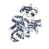
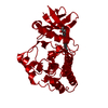
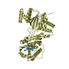
Entry Database : PDB / ID : 6crfTitle Crystal Structure of Shp2 E76K GOF Mutant in the Open Conformation Tyrosine-protein phosphatase non-receptor type 11 Keywords / / / / / / / Function / homology Function Domain/homology Component
/ / / / / / / / / / / / / / / / / / / / / / / / / / / / / / / / / / / / / / / / / / / / / / / / / / / / / / / / / / / / / / / / / / / / / / / / / / / / / / / / / / / / / / / / / / / / / / / / / / / / / / / / / / / / / / / / / / / / / / / / / / / / / / / / / / / / / / / / / / / / Biological species Homo sapiens (human)Method / / / Resolution : 2.62 Å Authors Stams, T. / Fodor, M. Journal : Nat Commun / Year : 2018Title : Structural reorganization of SHP2 by oncogenic mutations and implications for oncoprotein resistance to allosteric inhibition.Authors : LaRochelle, J.R. / Fodor, M. / Vemulapalli, V. / Mohseni, M. / Wang, P. / Stams, T. / LaMarche, M.J. / Chopra, R. / Acker, M.G. / Blacklow, S.C. History Deposition Mar 17, 2018 Deposition site / Processing site Revision 1.0 Nov 7, 2018 Provider / Type Revision 1.1 Oct 4, 2023 Group / Database references / Refinement descriptionCategory chem_comp_atom / chem_comp_bond ... chem_comp_atom / chem_comp_bond / database_2 / pdbx_initial_refinement_model / software Item / _database_2.pdbx_database_accession / _software.name
Show all Show less
 Movie
Movie Controller

 Yorodumi
 Open data
 Basic information
 Components
 Keywords
 Function and homology information
 Homo sapiens (human)
 X-RAY DIFFRACTION /
  Authors
 Citation
 Structure visualization
 Downloads & links
 Other downloads
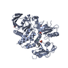


 PDBj
PDBj


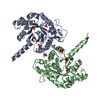
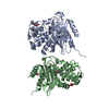
 Assembly


 Mass: 92.094 Da / Num. of mol.: 1 / Source method: obtained synthetically / Formula: C3H8O3
 Mass: 18.015 Da / Num. of mol.: 208 / Source method: isolated from a natural source / Formula: H2O
 Sample preparation
 / Beamline: 17-ID / Wavelength: 1 Å
 Processing