 Movie
Movie Controller

[English] 日本語
 Yorodumi
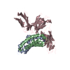
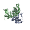
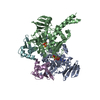
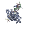
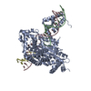
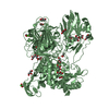
- PDB-6c83: Structure of Aurora A (122-403) bound to inhibitory Monobody Mb2 ... -
+ Open data

- Basic information
| Entry | Database: PDB / ID: 6c83 | ||||||||||||
|---|---|---|---|---|---|---|---|---|---|---|---|---|---|
| Title | Structure of Aurora A (122-403) bound to inhibitory Monobody Mb2 and AMPPCP | ||||||||||||
 Components Components |
| ||||||||||||
 Keywords Keywords | transferase/inhibitor / Transferase / Aurora A / Monobody / AMPPCP / Kinase / Activation / Allostery / Cell cycle / Cancer / transferase-inhibitor complex | ||||||||||||
| Function / homology |  Function and homology information Function and homology informationInteraction between PHLDA1 and AURKA / regulation of centrosome cycle / axon hillock / spindle assembly involved in female meiosis I / cilium disassembly / spindle pole centrosome / mitotic centrosome separation / histone H3S10 kinase activity / chromosome passenger complex / positive regulation of oocyte maturation ...Interaction between PHLDA1 and AURKA / regulation of centrosome cycle / axon hillock / spindle assembly involved in female meiosis I / cilium disassembly / spindle pole centrosome / mitotic centrosome separation / histone H3S10 kinase activity / chromosome passenger complex / positive regulation of oocyte maturation / pronucleus / germinal vesicle / protein localization to centrosome / meiotic spindle / anterior/posterior axis specification / neuron projection extension / centrosome localization / spindle organization / positive regulation of mitochondrial fission / mitotic spindle pole / spindle midzone / SUMOylation of DNA replication proteins / negative regulation of protein binding / regulation of G2/M transition of mitotic cell cycle / positive regulation of mitotic cell cycle / positive regulation of mitotic nuclear division / protein serine/threonine/tyrosine kinase activity / centriole / TP53 Regulates Transcription of Genes Involved in G2 Cell Cycle Arrest / liver regeneration / AURKA Activation by TPX2 / molecular function activator activity / regulation of signal transduction by p53 class mediator / mitotic spindle organization / peptidyl-serine phosphorylation / regulation of cytokinesis / regulation of protein stability / response to wounding / APC/C:Cdh1 mediated degradation of Cdc20 and other APC/C:Cdh1 targeted proteins in late mitosis/early G1 / FBXL7 down-regulates AURKA during mitotic entry and in early mitosis / G2/M transition of mitotic cell cycle / kinetochore / spindle / spindle pole / mitotic spindle / Regulation of PLK1 Activity at G2/M Transition / protein autophosphorylation / mitotic cell cycle / positive regulation of proteasomal ubiquitin-dependent protein catabolic process / microtubule cytoskeleton / midbody / Regulation of TP53 Activity through Phosphorylation / basolateral plasma membrane / microtubule / proteasome-mediated ubiquitin-dependent protein catabolic process / protein phosphorylation / protein kinase activity / non-specific serine/threonine protein kinase / ciliary basal body / postsynaptic density / protein heterodimerization activity / negative regulation of gene expression / protein serine kinase activity / cell division / protein serine/threonine kinase activity / apoptotic process / centrosome / ubiquitin protein ligase binding / protein kinase binding / negative regulation of apoptotic process / perinuclear region of cytoplasm / glutamatergic synapse / nucleoplasm / ATP binding / nucleus / cytosol Similarity search - Function | ||||||||||||
| Biological species |  Homo sapiens (human) Homo sapiens (human)synthetic construct (others) | ||||||||||||
| Method |  X-RAY DIFFRACTION / SYNCHROTRON / MOLECULAR REPLACEMENT / Resolution: 2.55 Å X-RAY DIFFRACTION / SYNCHROTRON / MOLECULAR REPLACEMENT / Resolution: 2.55 Å | ||||||||||||
 Authors Authors | Hoemberger, M. / Kutter, S. / Zorba, A. / Nguyen, V. / Shohei, A. / Shohei, K. / Kern, D. | ||||||||||||
| Funding support |  United States, 3items United States, 3items
| ||||||||||||
 Citation Citation | Journal: Proc.Natl.Acad.Sci.USA / Year: 2019 Title: Allosteric modulation of a human protein kinase with monobodies. Authors: Zorba, A. / Nguyen, V. / Koide, A. / Hoemberger, M. / Zheng, Y. / Kutter, S. / Kim, C. / Koide, S. / Kern, D. | ||||||||||||
| History |
|
- Structure visualization
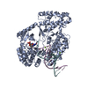
| Structure viewer | Molecule: MolmilJmol/JSmol |
|---|
- Downloads & links
-Download
| PDBx/mmCIF format | 6c83.cif.gz | 302.8 KB | Display | PDBx/mmCIF format |
|---|---|---|---|---|
| PDB format | pdb6c83.ent.gz | 211 KB | Display | PDB format |
| PDBx/mmJSON format | 6c83.json.gz | Tree view | PDBx/mmJSON format | |
| Others |  Other downloads Other downloads |
-Validation report
| Arichive directory | https://data.pdbj.org/pub/pdb/validation_reports/c8/6c83ftp://data.pdbj.org/pub/pdb/validation_reports/c8/6c83 | HTTPS FTP |
|---|
-Related structure data
| Related structure data | 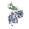 5g15C  3k2mS 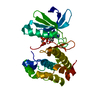 4c3rS S: Starting model for refinement C: citing same article ( |
|---|---|
| Similar structure data |

-Links
 PDBj
PDBj


- Assembly
| Deposited unit | 
| ||||||||||||
|---|---|---|---|---|---|---|---|---|---|---|---|---|---|
| 1 |
| ||||||||||||
| Unit cell |
|
-Components
| #1: Protein | Mass: 32990.754 Da / Num. of mol.: 2 Source method: isolated from a genetically manipulated source Source: (gene. exp.) Homo sapiens (human)Gene: AURKA, AIK, AIRK1, ARK1, AURA, AYK1, BTAK, IAK1, STK15, STK6 Production host:  References: UniProt: O14965, non-specific serine/threonine protein kinase #2: Protein | Mass: 9853.987 Da / Num. of mol.: 2 Source method: isolated from a genetically manipulated source Source: (gene. exp.) synthetic construct (others) / Production host: #3: Chemical |   Mass: 505.208 Da / Num. of mol.: 2 / Source method: obtained synthetically / Formula: C11H18N5O12P3 / Comment: AMP-PCP, energy-carrying molecule analogue*YM Mass: 505.208 Da / Num. of mol.: 2 / Source method: obtained synthetically / Formula: C11H18N5O12P3 / Comment: AMP-PCP, energy-carrying molecule analogue*YM |
|---|
-Experimental details
-Experiment
| Experiment | Method: X-RAY DIFFRACTION / Number of used crystals: 1 |
|---|
- Sample preparation
| Crystal | Density Matthews: 2.23 Å3/Da / Density % sol: 44.94 % |
|---|---|
| Crystal grow | Temperature: 291.15 K / Method: vapor diffusion, sitting drop / pH: 6.5 Details: Crystals of dephosphorylated Aurora A (122-403) in complex with Monobody Mb2 and AMPPCP were obtained by mixing a 1:1 ratio of 0.24mM Aurora A, 0.25mM Mb2 and 2.5mM AMPPCP in 20mM TrisHCl pH ...Details: Crystals of dephosphorylated Aurora A (122-403) in complex with Monobody Mb2 and AMPPCP were obtained by mixing a 1:1 ratio of 0.24mM Aurora A, 0.25mM Mb2 and 2.5mM AMPPCP in 20mM TrisHCl pH 7.5, 200mM NaCl,20mM MgCl2, 10% glycerol, 5mM TCEP with mother liquor (0.1M Bis-Tris pH 5.5, 0.2M NaCl, 25% (w/v) PEG 3350 |
-Data collection
| Diffraction | Mean temperature: 100 K |
|---|---|
| Diffraction source | Source: SYNCHROTRON / Site: SSRL / Beamline: BL7-1 / Wavelength: 0.9795 Å |
| Detector | Type: ADSC QUANTUM 315r / Detector: CCD / Date: Jan 28, 2017 |
| Radiation | Monochromator: Si(111) side scattering I-beam bent single crystal Protocol: SINGLE WAVELENGTH / Monochromatic (M) / Laue (L): M / Scattering type: x-ray |
| Radiation wavelength | Wavelength: 0.9795 Å / Relative weight: 1 |
| Reflection | Resolution: 2.55→64.86 Å / Num. obs: 25451 / % possible obs: 98.47 % / Redundancy: 5.3 % / Biso Wilson estimate: 62.83 Å2 / Rmerge(I) obs: 0.053 / Net I/σ(I): 13.1 |
| Reflection shell | Resolution: 2.55→2.62 Å |
- Processing
| Software |
| |||||||||||||||||||||||||||||||||||||||||||||||||||||||||||||||||||||||||||||||||||||||||||||||||||||||||
|---|---|---|---|---|---|---|---|---|---|---|---|---|---|---|---|---|---|---|---|---|---|---|---|---|---|---|---|---|---|---|---|---|---|---|---|---|---|---|---|---|---|---|---|---|---|---|---|---|---|---|---|---|---|---|---|---|---|---|---|---|---|---|---|---|---|---|---|---|---|---|---|---|---|---|---|---|---|---|---|---|---|---|---|---|---|---|---|---|---|---|---|---|---|---|---|---|---|---|---|---|---|---|---|---|---|---|
| Refinement | Method to determine structure: MOLECULAR REPLACEMENT Starting model: 4c3r, 3k2m Resolution: 2.55→46.82 Å / SU ML: 0.4642 / Cross valid method: FREE R-VALUE / σ(F): 1.35 / Phase error: 37.2497
| |||||||||||||||||||||||||||||||||||||||||||||||||||||||||||||||||||||||||||||||||||||||||||||||||||||||||
| Solvent computation | Shrinkage radii: 0.9 Å / VDW probe radii: 1.11 Å | |||||||||||||||||||||||||||||||||||||||||||||||||||||||||||||||||||||||||||||||||||||||||||||||||||||||||
| Displacement parameters | Biso mean: 73.75 Å2 | |||||||||||||||||||||||||||||||||||||||||||||||||||||||||||||||||||||||||||||||||||||||||||||||||||||||||
| Refinement step | Cycle: LAST / Resolution: 2.55→46.82 Å
| |||||||||||||||||||||||||||||||||||||||||||||||||||||||||||||||||||||||||||||||||||||||||||||||||||||||||
| Refine LS restraints |
| |||||||||||||||||||||||||||||||||||||||||||||||||||||||||||||||||||||||||||||||||||||||||||||||||||||||||
| LS refinement shell |
|
