 Movie
Movie Controller

[English] 日本語
 Yorodumi
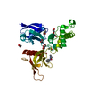
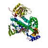
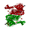
- PDB-5g15: Structure Aurora A (122-403) bound to activating monobody Mb1 and... -
+ Open data

- Basic information
| Entry | Database: PDB / ID: 5g15 | ||||||
|---|---|---|---|---|---|---|---|
| Title | Structure Aurora A (122-403) bound to activating monobody Mb1 and AMPPCP | ||||||
 Components Components |
| ||||||
 Keywords Keywords | TRANSFERASE / AURORA A / MONOBODY / AMPPCP / KINASE / ACTIVATION / ALLOSTERY / CELL CYCLE / CANCER | ||||||
| Function / homology |  Function and homology information Function and homology informationInteraction between PHLDA1 and AURKA / regulation of centrosome cycle / axon hillock / spindle assembly involved in female meiosis I / cilium disassembly / spindle pole centrosome / mitotic centrosome separation / histone H3S10 kinase activity / chromosome passenger complex / positive regulation of oocyte maturation ...Interaction between PHLDA1 and AURKA / regulation of centrosome cycle / axon hillock / spindle assembly involved in female meiosis I / cilium disassembly / spindle pole centrosome / mitotic centrosome separation / histone H3S10 kinase activity / chromosome passenger complex / positive regulation of oocyte maturation / pronucleus / germinal vesicle / protein localization to centrosome / meiotic spindle / anterior/posterior axis specification / neuron projection extension / centrosome localization / spindle organization / positive regulation of mitochondrial fission / mitotic spindle pole / spindle midzone / SUMOylation of DNA replication proteins / negative regulation of protein binding / regulation of G2/M transition of mitotic cell cycle / positive regulation of mitotic cell cycle / positive regulation of mitotic nuclear division / protein serine/threonine/tyrosine kinase activity / centriole / TP53 Regulates Transcription of Genes Involved in G2 Cell Cycle Arrest / liver regeneration / AURKA Activation by TPX2 / molecular function activator activity / regulation of signal transduction by p53 class mediator / mitotic spindle organization / peptidyl-serine phosphorylation / regulation of cytokinesis / regulation of protein stability / response to wounding / APC/C:Cdh1 mediated degradation of Cdc20 and other APC/C:Cdh1 targeted proteins in late mitosis/early G1 / FBXL7 down-regulates AURKA during mitotic entry and in early mitosis / G2/M transition of mitotic cell cycle / kinetochore / spindle / spindle pole / mitotic spindle / Regulation of PLK1 Activity at G2/M Transition / protein autophosphorylation / mitotic cell cycle / positive regulation of proteasomal ubiquitin-dependent protein catabolic process / microtubule cytoskeleton / midbody / Regulation of TP53 Activity through Phosphorylation / basolateral plasma membrane / microtubule / proteasome-mediated ubiquitin-dependent protein catabolic process / protein phosphorylation / protein kinase activity / non-specific serine/threonine protein kinase / ciliary basal body / postsynaptic density / protein heterodimerization activity / negative regulation of gene expression / protein serine kinase activity / cell division / protein serine/threonine kinase activity / apoptotic process / centrosome / ubiquitin protein ligase binding / protein kinase binding / negative regulation of apoptotic process / perinuclear region of cytoplasm / glutamatergic synapse / nucleoplasm / ATP binding / nucleus / cytosol Similarity search - Function | ||||||
| Biological species |  HOMO SAPIENS (human) HOMO SAPIENS (human)SYNTHETIC CONSTRUCT (others) | ||||||
| Method |  X-RAY DIFFRACTION / SYNCHROTRON / MOLECULAR REPLACEMENT / Resolution: 2.06 Å X-RAY DIFFRACTION / SYNCHROTRON / MOLECULAR REPLACEMENT / Resolution: 2.06 Å | ||||||
 Authors Authors | Zorba, A. / Kutter, S. / Kern, D. / Koide, S. / Koide, A. | ||||||
 Citation Citation | Journal: Proc.Natl.Acad.Sci.USA / Year: 2019 Title: Allosteric modulation of a human protein kinase with monobodies. Authors: Zorba, A. / Nguyen, V. / Koide, A. / Hoemberger, M. / Zheng, Y. / Kutter, S. / Kim, C. / Koide, S. / Kern, D. | ||||||
| History |
|
- Structure visualization
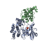
| Structure viewer | Molecule: MolmilJmol/JSmol |
|---|
- Downloads & links
-Download
| PDBx/mmCIF format | 5g15.cif.gz | 92.8 KB | Display | PDBx/mmCIF format |
|---|---|---|---|---|
| PDB format | pdb5g15.ent.gz | 69.4 KB | Display | PDB format |
| PDBx/mmJSON format | 5g15.json.gz | Tree view | PDBx/mmJSON format | |
| Others |  Other downloads Other downloads |
-Validation report
| Arichive directory | https://data.pdbj.org/pub/pdb/validation_reports/g1/5g15ftp://data.pdbj.org/pub/pdb/validation_reports/g1/5g15 | HTTPS FTP |
|---|
-Related structure data
| Related structure data | 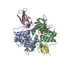 6c83C  3k2mS 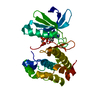 4c3rS  5g16 S: Starting model for refinement C: citing same article ( |
|---|---|
| Similar structure data |


-Links
 PDBj
PDBj


- Assembly
| Deposited unit | 
| ||||||||
|---|---|---|---|---|---|---|---|---|---|
| 1 |
| ||||||||
| Unit cell |
| ||||||||
| Components on special symmetry positions |
|
-Components
-Protein / Antibody , 2 types, 2 molecules AB
| #1: Protein | Mass: 32689.459 Da / Num. of mol.: 1 / Fragment: KINASE DOMAIN, UNP RESIDUES 122-403 Source method: isolated from a genetically manipulated source Source: (gene. exp.) HOMO SAPIENS (human) / Plasmid: PET28A / Production host:  References: UniProt: O14965, non-specific serine/threonine protein kinase |
|---|---|
| #2: Antibody | Mass: 10577.592 Da / Num. of mol.: 1 Source method: isolated from a genetically manipulated source Source: (gene. exp.) SYNTHETIC CONSTRUCT (others) / Plasmid: PET28A / Production host: |
-Non-polymers , 4 types, 151 molecules 


| #3: Chemical | ChemComp-ACP /  Mass: 505.208 Da / Num. of mol.: 1 / Source method: obtained synthetically / Formula: C11H18N5O12P3 / Comment: AMP-PCP, energy-carrying molecule analogue*YM Mass: 505.208 Da / Num. of mol.: 1 / Source method: obtained synthetically / Formula: C11H18N5O12P3 / Comment: AMP-PCP, energy-carrying molecule analogue*YM | ||
|---|---|---|---|
| #4: Chemical | ChemComp-MG /  Mass: 24.305 Da / Num. of mol.: 1 / Source method: obtained synthetically / Formula: Mg Mass: 24.305 Da / Num. of mol.: 1 / Source method: obtained synthetically / Formula: Mg | ||
| #5: Chemical | ChemComp-SO4 /  Mass: 96.063 Da / Num. of mol.: 4 / Source method: obtained synthetically / Formula: SO4 Mass: 96.063 Da / Num. of mol.: 4 / Source method: obtained synthetically / Formula: SO4#6: Water | ChemComp-HOH / | Mass: 18.015 Da / Num. of mol.: 145 / Source method: isolated from a natural source / Formula: H2O |
-Experimental details
-Experiment
| Experiment | Method: X-RAY DIFFRACTION / Number of used crystals: 1 |
|---|
- Sample preparation
| Crystal | Density Matthews: 2.86 Å3/Da / Density % sol: 57.04 % / Description: NONE |
|---|---|
| Crystal grow | Temperature: 291 K / Method: vapor diffusion, sitting drop / pH: 6.5 Details: AURORA A AND MONOBODIES WERE ALIQUOTED IN STORAGE BUFFER (20MM TRISHCL, 200MM NACL, 10% (V/V) GLYCEROL, 20MM MGCL2, 5MM TCEP, PH 7.50) AND KEPT AT -80C. AMPPCP WAS PREPARED FRESH FROM POWDER ...Details: AURORA A AND MONOBODIES WERE ALIQUOTED IN STORAGE BUFFER (20MM TRISHCL, 200MM NACL, 10% (V/V) GLYCEROL, 20MM MGCL2, 5MM TCEP, PH 7.50) AND KEPT AT -80C. AMPPCP WAS PREPARED FRESH FROM POWDER THE DAY OF CRYSTALLIZATION IN CONCENTRATIONS OF 100-120MM IN STORAGE BUFFER. CRYSTALS OF AURA IN COMPLEX WITH AMPPCP AND ACTIVATING MONOBODY, MB1, WERE OBTAINED BY COMBINING 0.5UL OF [300UM AURA WITH 5MM AMPPCP AND 300UM MB1] WITH 0.5UL OF MOTHER LIQUOR (0.1M MES SODIUM SALT PH 6.50, 0.2M AMMONIUM SULFATE, 4% (V/V) 1,3-PROPANEDIOL, 30% (W/V) PEG8000). CRYSTALS WERE GROWN AT 18C BY VAPOR DIFFUSION AND THE SITTING DROP METHOD. THE CRYSTALS WERE WASHED WITH MOTHER LIQUOR AND FLASH FROZEN IN LIQUID NITROGEN IN PREPARATION FOR DATA COLLECTION. |
-Data collection
| Diffraction | Mean temperature: 100 K |
|---|---|
| Diffraction source | Source: SYNCHROTRON / Site: ALS  / Beamline: 8.2.2 / Wavelength: 0.99992 / Beamline: 8.2.2 / Wavelength: 0.99992 |
| Detector | Type: ADSC QUANTUM 315 / Detector: CCD / Date: May 9, 2015 |
| Radiation | Monochromator: DOUBLE-CRYSTAL SI(111) / Protocol: SINGLE WAVELENGTH / Monochromatic (M) / Laue (L): M / Scattering type: x-ray |
| Radiation wavelength | Wavelength: 0.99992 Å / Relative weight: 1 |
| Reflection | Resolution: 2.06→58.13 Å / Num. obs: 30879 / % possible obs: 100 % / Observed criterion σ(I): 1.8 / Redundancy: 7.2 % / Rmerge(I) obs: 0.13 / Net I/σ(I): 10.3 |
| Reflection shell | Resolution: 2.06→2.12 Å / Redundancy: 7.2 % / Rmerge(I) obs: 1.21 / Mean I/σ(I) obs: 1.8 / % possible all: 99.5 |
- Processing
| Software |
| ||||||||||||||||||||||||||||||||||||||||||||||||||||||||||||||||||||||||||||||||||||||||||||||||||||||||||||||||||||||||||||||||||||||||||||||||||||||||||||||||||||||||||||||||||||||
|---|---|---|---|---|---|---|---|---|---|---|---|---|---|---|---|---|---|---|---|---|---|---|---|---|---|---|---|---|---|---|---|---|---|---|---|---|---|---|---|---|---|---|---|---|---|---|---|---|---|---|---|---|---|---|---|---|---|---|---|---|---|---|---|---|---|---|---|---|---|---|---|---|---|---|---|---|---|---|---|---|---|---|---|---|---|---|---|---|---|---|---|---|---|---|---|---|---|---|---|---|---|---|---|---|---|---|---|---|---|---|---|---|---|---|---|---|---|---|---|---|---|---|---|---|---|---|---|---|---|---|---|---|---|---|---|---|---|---|---|---|---|---|---|---|---|---|---|---|---|---|---|---|---|---|---|---|---|---|---|---|---|---|---|---|---|---|---|---|---|---|---|---|---|---|---|---|---|---|---|---|---|---|---|
| Refinement | Method to determine structure: MOLECULAR REPLACEMENT Starting model: PDB ENTRIES 4C3R AND 3K2M Resolution: 2.06→77.1 Å / Cor.coef. Fo:Fc: 0.937 / Cor.coef. Fo:Fc free: 0.912 / SU B: 7.946 / SU ML: 0.185 / Cross valid method: THROUGHOUT / ESU R: 0.2 / ESU R Free: 0.184 / Stereochemistry target values: MAXIMUM LIKELIHOOD / Details: HYDROGENS HAVE BEEN ADDED IN THE RIDING POSITIONS.
| ||||||||||||||||||||||||||||||||||||||||||||||||||||||||||||||||||||||||||||||||||||||||||||||||||||||||||||||||||||||||||||||||||||||||||||||||||||||||||||||||||||||||||||||||||||||
| Solvent computation | Ion probe radii: 0.8 Å / Shrinkage radii: 0.8 Å / VDW probe radii: 1.2 Å / Solvent model: MASK | ||||||||||||||||||||||||||||||||||||||||||||||||||||||||||||||||||||||||||||||||||||||||||||||||||||||||||||||||||||||||||||||||||||||||||||||||||||||||||||||||||||||||||||||||||||||
| Displacement parameters | Biso mean: 34.133 Å2
| ||||||||||||||||||||||||||||||||||||||||||||||||||||||||||||||||||||||||||||||||||||||||||||||||||||||||||||||||||||||||||||||||||||||||||||||||||||||||||||||||||||||||||||||||||||||
| Refinement step | Cycle: LAST / Resolution: 2.06→77.1 Å
| ||||||||||||||||||||||||||||||||||||||||||||||||||||||||||||||||||||||||||||||||||||||||||||||||||||||||||||||||||||||||||||||||||||||||||||||||||||||||||||||||||||||||||||||||||||||
| Refine LS restraints |
|