- PDB-4c3r: Structure of dephosphorylated Aurora A (122-403) bound to AMPPCP -
+
Open data
ID or keywords:
Loading...
-
Basic information
Entry
Database: PDB / ID: 4c3r
Title
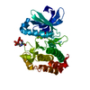
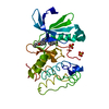
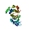
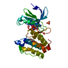
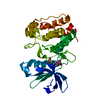
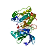
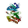
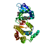
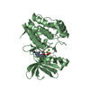
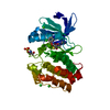
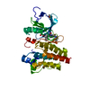
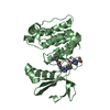
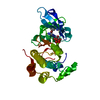
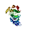
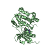
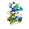
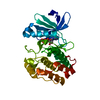
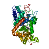
Structure of dephosphorylated Aurora A (122-403) bound to AMPPCP
Components
AURORA KINASE A
Keywords
TRANSFERASE / AURORA A / ACTIVATION / CELL CYCLE / CANCER
Function / homology
Function and homology information
Interaction between PHLDA1 and AURKA / regulation of centrosome cycle / axon hillock / spindle assembly involved in female meiosis I / cilium disassembly / spindle pole centrosome / mitotic centrosome separation / histone H3S10 kinase activity / chromosome passenger complex / positive regulation of oocyte maturation ...Interaction between PHLDA1 and AURKA / regulation of centrosome cycle / axon hillock / spindle assembly involved in female meiosis I / cilium disassembly / spindle pole centrosome / mitotic centrosome separation / histone H3S10 kinase activity / chromosome passenger complex / positive regulation of oocyte maturation / pronucleus / germinal vesicle / protein localization to centrosome / meiotic spindle / anterior/posterior axis specification / neuron projection extension / centrosome localization / spindle organization / positive regulation of mitochondrial fission / mitotic spindle pole / spindle midzone / SUMOylation of DNA replication proteins / negative regulation of protein binding / regulation of G2/M transition of mitotic cell cycle / positive regulation of mitotic cell cycle / positive regulation of mitotic nuclear division / protein serine/threonine/tyrosine kinase activity / centriole / TP53 Regulates Transcription of Genes Involved in G2 Cell Cycle Arrest / liver regeneration / AURKA Activation by TPX2 / molecular function activator activity / regulation of signal transduction by p53 class mediator / mitotic spindle organization / peptidyl-serine phosphorylation / regulation of cytokinesis / regulation of protein stability / response to wounding / APC/C:Cdh1 mediated degradation of Cdc20 and other APC/C:Cdh1 targeted proteins in late mitosis/early G1 / FBXL7 down-regulates AURKA during mitotic entry and in early mitosis / G2/M transition of mitotic cell cycle / kinetochore / spindle / spindle pole / mitotic spindle / Regulation of PLK1 Activity at G2/M Transition / protein autophosphorylation / mitotic cell cycle / positive regulation of proteasomal ubiquitin-dependent protein catabolic process / microtubule cytoskeleton / midbody / Regulation of TP53 Activity through Phosphorylation / basolateral plasma membrane / microtubule / proteasome-mediated ubiquitin-dependent protein catabolic process / protein phosphorylation / protein kinase activity / non-specific serine/threonine protein kinase / ciliary basal body / postsynaptic density / protein heterodimerization activity / negative regulation of gene expression / protein serine kinase activity / cell division / protein serine/threonine kinase activity / apoptotic process / centrosome / ubiquitin protein ligase binding / protein kinase binding / negative regulation of apoptotic process / perinuclear region of cytoplasm / glutamatergic synapse / nucleoplasm / ATP binding / nucleus / cytosol Similarity search - Function
Aurora kinase A / Aurora kinase / Phosphorylase Kinase; domain 1 / Phosphorylase Kinase; domain 1 / Transferase(Phosphotransferase) domain 1 / Transferase(Phosphotransferase); domain 1 / Serine/threonine-protein kinase, active site / Serine/Threonine protein kinases active-site signature. / Protein kinase domain / Serine/Threonine protein kinases, catalytic domain ...Aurora kinase A / Aurora kinase / Phosphorylase Kinase; domain 1 / Phosphorylase Kinase; domain 1 / Transferase(Phosphotransferase) domain 1 / Transferase(Phosphotransferase); domain 1 / Serine/threonine-protein kinase, active site / Serine/Threonine protein kinases active-site signature. / Protein kinase domain / Serine/Threonine protein kinases, catalytic domain / Protein kinase, ATP binding site / Protein kinases ATP-binding region signature. / Protein kinase domain profile. / Protein kinase domain / Protein kinase-like domain superfamily / 2-Layer Sandwich / Orthogonal Bundle / Mainly Alpha / Alpha Beta Similarity search - Domain/homology
Method: X-RAY DIFFRACTION / Number of used crystals: 1
-
Sample preparation
Crystal
Density Matthews: 2.96 Å3/Da / Density % sol: 58.54 % / Description: NONE
Crystal grow
Temperature: 291 K / Method: vapor diffusion, hanging drop / pH: 7.5 Details: CRYSTALS OF DEPHOSPHORYLATED A(122-403) IN COMPLEX WITH AMPPCP WERE OBTAINED BY MIXING A 2:1 RATIO OF 570UM (18MG/ML) DEP A(122-403) AND 1MM AMPPCP WITH MOTHER LIQUOR (0.2M AMMONIUM SULFATE, ...Details: CRYSTALS OF DEPHOSPHORYLATED A(122-403) IN COMPLEX WITH AMPPCP WERE OBTAINED BY MIXING A 2:1 RATIO OF 570UM (18MG/ML) DEP A(122-403) AND 1MM AMPPCP WITH MOTHER LIQUOR (0.2M AMMONIUM SULFATE, 0.2M TRISHCL PH 7.50, 30% (W/V) PEG3350). THE CRYSTALS WERE GROWN AT 18C BY VAPOR DIFFUSION AND THE HANGING DROP METHOD.
Resolution: 2.79→55.42 Å / Cor.coef. Fo:Fc: 0.944 / Cor.coef. Fo:Fc free: 0.901 / SU B: 16.737 / SU ML: 0.332 / Cross valid method: THROUGHOUT / ESU R: 4.704 / ESU R Free: 0.442 / Stereochemistry target values: MAXIMUM LIKELIHOOD / Details: HYDROGENS HAVE BEEN ADDED IN THE RIDING POSITIONS.
Rfactor
Num. reflection
% reflection
Selection details
Rfree
0.30601
941
10 %
RANDOM
Rwork
0.22124
-
-
-
obs
0.22939
8459
99.97 %
-
Solvent computation
Ion probe radii: 0.8 Å / Shrinkage radii: 0.8 Å / VDW probe radii: 1.2 Å / Solvent model: MASK
 Movie
Movie Controller


 Open data
 Basic information
 Components
 Keywords
 Function and homology information
 HOMO SAPIENS (human)
 X-RAY DIFFRACTION /
  Authors
 Citation
 Structure visualization
 Downloads & links
 Other downloads
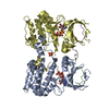


 PDBj
PDBj


 Assembly
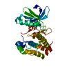


 Mass: 505.208 Da / Num. of mol.: 1 / Source method: obtained synthetically / Formula: C11H18N5O12P3 / Comment: AMP-PCP, energy-carrying molecule analogue*YM
 Sample preparation
 / Beamline: 8.2.2 / Wavelength: 0.999941
  Processing