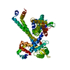
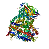
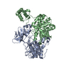
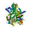
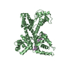
- PDB-5afq: Crystal structure of RPC62 - RPC32 beta -
+
Open data
ID or keywords:
Loading...
-
Basic information
Entry
Database: PDB / ID: 5afq
Title
Crystal structure of RPC62 - RPC32 beta
Components
DNA-DIRECTED RNA POLYMERASE III SUBUNIT RPC3
RPC32 BETA (RPC7L)
Keywords
REPLICATION / HUMAN RNA POLYMERASE III
Function / homology
Function and homology information
RNA Polymerase III Chain Elongation / RNA Polymerase III Transcription Termination / regulation of transcription by RNA polymerase III / RNA Polymerase III Transcription Initiation From Type 1 Promoter / RNA Polymerase III Transcription Initiation From Type 2 Promoter / RNA Polymerase III Transcription Initiation From Type 3 Promoter / RNA Polymerase III Abortive And Retractive Initiation / Cytosolic sensors of pathogen-associated DNA / positive regulation of innate immune response / RNA polymerase III complex ...RNA Polymerase III Chain Elongation / RNA Polymerase III Transcription Termination / regulation of transcription by RNA polymerase III / RNA Polymerase III Transcription Initiation From Type 1 Promoter / RNA Polymerase III Transcription Initiation From Type 2 Promoter / RNA Polymerase III Transcription Initiation From Type 3 Promoter / RNA Polymerase III Abortive And Retractive Initiation / Cytosolic sensors of pathogen-associated DNA / positive regulation of innate immune response / RNA polymerase III complex / positive regulation of interferon-beta production / DNA-directed RNA polymerase activity / single-stranded DNA binding / defense response to virus / innate immune response / DNA-templated transcription / nucleoplasm / cytosol Similarity search - Function
RNA polymerase III Rpc82, C -terminal / DNA-directed RNA polymerase III subunit RPC3 / : / RNA polymerase III subunit RPC82 / DNA-directed RNA polymerase III subunit RPC3, helical hairpin domain / POLR3C, C-terminal winged-helix domain / RNA polymerase III subunit RPC82-related, helix-turn-helix / RNA polymerase III subunit RPC82 helix-turn-helix domain / Winged helix-like DNA-binding domain superfamily Similarity search - Domain/homology
DNA-DIRECTEDRNAPOLYMERASEIIISUBUNITRPC3 / RNA POLYMERASE III SUBUNIT C3 / DNA-DIRECTED RNA POLYMERASE III SUBUNIT C / RNA POLYMERASE III 62 ...RNA POLYMERASE III SUBUNIT C3 / DNA-DIRECTED RNA POLYMERASE III SUBUNIT C / RNA POLYMERASE III 62 KDA SUBUNIT / RPC62 / RPC62
Mass: 60692.555 Da / Num. of mol.: 2 Source method: isolated from a genetically manipulated source Source: (gene. exp.) HOMO SAPIENS (human) / Production host: ESCHERICHIA COLI (E. coli) / Strain (production host): BL21(DE3) / Variant (production host): ROSETTA / References: UniProt: Q9BUI4
#2: Protein
RPC32BETA (RPC7L)
Mass: 18570.826 Da / Num. of mol.: 2 Source method: isolated from a genetically manipulated source Source: (gene. exp.) HOMO SAPIENS (human) / Production host: ESCHERICHIA COLI (E. coli) / Strain (production host): BL21(DE3) / Variant (production host): ROSETTA
Sequence details
CHAIN D AND E IS RPC32 BETA (RPC7L), FRAGMENT 50-134 EXPRESSED AS RPC62 IN ROSETTA (DE3) STRAINS ...CHAIN D AND E IS RPC32 BETA (RPC7L), FRAGMENT 50-134 EXPRESSED AS RPC62 IN ROSETTA (DE3) STRAINS FROM E. COLI. SINCE THE REGISTER OF THE AMINO ACIDS ARE NOT KNOWN, CHAINS D AND E HAS BEEN BUILT IN AS UNK IN THE MODEL.
-
Experimental details
-
Experiment
Experiment
Method: X-RAY DIFFRACTION / Number of used crystals: 1
-
Sample preparation
Crystal
Density Matthews: 3.89 Å3/Da / Density % sol: 70 % / Description: NONE
In the structure databanks used in Yorodumi, some data are registered as the other names, "COVID-19 virus" and "2019-nCoV". Here are the details of the virus and the list of structure data.
Jan 31, 2019. EMDB accession codes are about to change! (news from PDBe EMDB page)
EMDB accession codes are about to change! (news from PDBe EMDB page)
The allocation of 4 digits for EMDB accession codes will soon come to an end. Whilst these codes will remain in use, new EMDB accession codes will include an additional digit and will expand incrementally as the available range of codes is exhausted. The current 4-digit format prefixed with “EMD-” (i.e. EMD-XXXX) will advance to a 5-digit format (i.e. EMD-XXXXX), and so on. It is currently estimated that the 4-digit codes will be depleted around Spring 2019, at which point the 5-digit format will come into force.
The EM Navigator/Yorodumi systems omit the EMD- prefix.
Related info.:Q: What is EMD? / ID/Accession-code notation in Yorodumi/EM Navigator
Yorodumi is a browser for structure data from EMDB, PDB, SASBDB, etc.
This page is also the successor to EM Navigator detail page, and also detail information page/front-end page for Omokage search.
The word "yorodu" (or yorozu) is an old Japanese word meaning "ten thousand". "mi" (miru) is to see.
Related info.:EMDB / PDB / SASBDB / Comparison of 3 databanks / Yorodumi Search / Aug 31, 2016. New EM Navigator & Yorodumi / Yorodumi Papers / Jmol/JSmol / Function and homology information / Changes in new EM Navigator and Yorodumi
 Movie
Movie Controller


 Open data
 Basic information
 Components
 Keywords
 Function and homology information
 HOMO SAPIENS (human)
 X-RAY DIFFRACTION /
  Authors
 Citation
 Structure visualization
 Downloads & links
 Other downloads


 PDBj
PDBj


 Assembly


 Sample preparation
 / Beamline: ID23-1 / Wavelength: 0.9763
  Processing