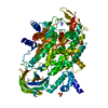
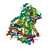
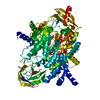
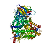
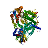
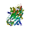
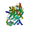
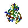
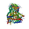
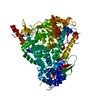
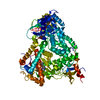
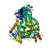
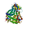
Entry Database : PDB / ID : 4flhTitle Crystal structure of human PI3K-gamma in complex with AMG511 Phosphatidylinositol 4,5-bisphosphate 3-kinase catalytic subunit gamma isoform Keywords / / / / / / Function / homology Function Domain/homology Component
/ / / / / / / / / / / / / / / / / / / / / / / / / / / / / / / / / / / / / / / / / / / / / / / / / / / / / / / / / / / / / / / / / / / / / / / / / / / / / / / / / / / / / / / / / / / / / / / / / / / / / / / / / / / / / / / / / / / / / / / / / / / / / / / / / / / / Biological species Homo sapiens (human)Method / / / Resolution : 2.6 Å Authors Whittington, D.A. / Tang, J. / Yakowec, P. Journal : J.Med.Chem. / Year : 2012Title : Selective Class I Phosphoinositide 3-Kinase Inhibitors: Optimization of a Series of Pyridyltriazines Leading to the Identification of a Clinical Candidate, AMG 511.Authors: Norman, M.H. / Andrews, K.L. / Bo, Y.Y. / Booker, S.K. / Caenepeel, S. / Cee, V.J. / D'Angelo, N.D. / Freeman, D.J. / Herberich, B.J. / Hong, F.T. / Jackson, C.L. / Jiang, J. / Lanman, B.A. ... Authors : Norman, M.H. / Andrews, K.L. / Bo, Y.Y. / Booker, S.K. / Caenepeel, S. / Cee, V.J. / D'Angelo, N.D. / Freeman, D.J. / Herberich, B.J. / Hong, F.T. / Jackson, C.L. / Jiang, J. / Lanman, B.A. / Liu, L. / McCarter, J.D. / Mullady, E.L. / Nishimura, N. / Pettus, L.H. / Reed, A.B. / Miguel, T.S. / Smith, A.L. / Stec, M.M. / Tadesse, S. / Tasker, A. / Aidasani, D. / Zhu, X. / Subramanian, R. / Tamayo, N.A. / Wang, L. / Whittington, D.A. / Wu, B. / Wu, T. / Wurz, R.P. / Yang, K. / Zalameda, L. / Zhang, N. / Hughes, P.E. History Deposition Jun 14, 2012 Deposition site / Processing site Revision 1.0 Aug 29, 2012 Provider / Type Revision 1.1 Oct 3, 2012 Group Revision 1.2 Nov 15, 2017 Group / Category Revision 1.3 Feb 28, 2024 Group / Database references / Derived calculationsCategory chem_comp_atom / chem_comp_bond ... chem_comp_atom / chem_comp_bond / database_2 / struct_ref_seq_dif / struct_site Item _database_2.pdbx_DOI / _database_2.pdbx_database_accession ... _database_2.pdbx_DOI / _database_2.pdbx_database_accession / _struct_ref_seq_dif.details / _struct_site.pdbx_auth_asym_id / _struct_site.pdbx_auth_comp_id / _struct_site.pdbx_auth_seq_id
Show all Show less
 Movie
Movie Controller


 Open data
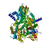
 Basic information
 Components
 Keywords
 Function and homology information
 Homo sapiens (human)
 X-RAY DIFFRACTION /
  Authors
 Citation
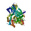
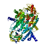
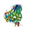
 Structure visualization
 Downloads & links
 Other downloads


 PDBj
PDBj


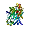
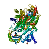
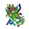
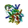
 Assembly
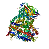

 Spodoptera frugiperda (fall armyworm)
 Mass: 96.063 Da / Num. of mol.: 4 / Source method: obtained synthetically / Formula: SO4
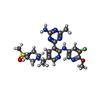
 Mass: 517.580 Da / Num. of mol.: 1 / Source method: obtained synthetically / Formula: C22H28FN9O3S
 Mass: 18.015 Da / Num. of mol.: 42 / Source method: isolated from a natural source / Formula: H2O
 Sample preparation
 / Beamline: 08ID-1 / Wavelength: 0.98 Å
 Processing