[English] 日本語
Yorodumi
- PDB-3r7r: Structure-based design of thienobenzoxepin inhibitors of PI3-Kinase -

+
Open data


ID or keywords:

Loading...

-
Basic information

Entry
Database: PDB / ID: 3r7r
TitleStructure-based design of thienobenzoxepin inhibitors of PI3-Kinase
ComponentsPhosphatidylinositol-4,5-bisphosphate 3-kinase catalytic subunit gamma isoform
KeywordsTRANSFERASE/TRANSFERASE INHIBITOR / kinase p110 / TRANSFERASE-TRANSFERASE INHIBITOR complex
Function / homology
Function and homology information


secretory granule localization / negative regulation of triglyceride catabolic process / natural killer cell chemotaxis / neutrophil extravasation / phosphatidylinositol-4-phosphate 3-kinase / regulation of calcium ion transmembrane transport / positive regulation of acute inflammatory response / respiratory burst involved in defense response / negative regulation of cardiac muscle contraction / T cell chemotaxis ...secretory granule localization / negative regulation of triglyceride catabolic process / natural killer cell chemotaxis / neutrophil extravasation / phosphatidylinositol-4-phosphate 3-kinase / regulation of calcium ion transmembrane transport / positive regulation of acute inflammatory response / respiratory burst involved in defense response / negative regulation of cardiac muscle contraction / T cell chemotaxis / negative regulation of fibroblast apoptotic process / phosphatidylinositol 3-kinase complex, class IB / sphingosine-1-phosphate receptor signaling pathway / dendritic cell chemotaxis / 1-phosphatidylinositol-4-phosphate 3-kinase activity / 1-phosphatidylinositol-4,5-bisphosphate 3-kinase activity / phosphatidylinositol-4,5-bisphosphate 3-kinase / phosphatidylinositol 3-kinase / phosphatidylinositol 3-kinase complex, class IA / phosphatidylinositol-3-phosphate biosynthetic process / 1-phosphatidylinositol-3-kinase activity / mast cell degranulation / Erythropoietin activates Phosphoinositide-3-kinase (PI3K) / hepatocyte apoptotic process / positive regulation of Rac protein signal transduction / regulation of cell adhesion mediated by integrin / Synthesis of PIPs at the plasma membrane / phosphatidylinositol phosphate biosynthetic process / regulation of angiogenesis / T cell proliferation / cellular response to cAMP / GPVI-mediated activation cascade / T cell activation / ephrin receptor binding / neutrophil chemotaxis / positive regulation of endothelial cell migration / phosphatidylinositol 3-kinase/protein kinase B signal transduction / positive regulation of cytokine production / positive regulation of MAP kinase activity / platelet aggregation / endocytosis / G beta:gamma signalling through PI3Kgamma / Signaling by CSF1 (M-CSF) in myeloid cells / kinase activity / positive regulation of cytosolic calcium ion concentration / angiogenesis / adaptive immune response / positive regulation of phosphatidylinositol 3-kinase/protein kinase B signal transduction / non-specific serine/threonine protein kinase / protein kinase activity / immune response / inflammatory response / G protein-coupled receptor signaling pathway / phosphorylation / protein serine kinase activity / innate immune response / protein serine/threonine kinase activity / ATP binding / membrane / identical protein binding / plasma membrane / cytosol / cytoplasm
Similarity search - Function
PIK3 catalytic subunit gamma, adaptor-binding domain / PIK3 catalytic subunit gamma adaptor-binding domain / Phosphatidylinositol 3-/4-kinase, catalytic domain / Phosphatidylinositol 3-kinase, accessory domain (PIK) / Phosphatidylinositol 3-kinase Catalytic Subunit; Chain A, domain 4 / Phosphatidylinositol 3-kinase Catalytic Subunit; Chain A, domain 4 / Phosphatidylinositol 3-kinase Catalytic Subunit; Chain A, Domain 5 / C2 domain / Phosphatidylinositol 3-kinase, adaptor-binding domain / Phosphatidylinositol 3-kinase adaptor-binding (PI3K ABD) domain profile. ...PIK3 catalytic subunit gamma, adaptor-binding domain / PIK3 catalytic subunit gamma adaptor-binding domain / Phosphatidylinositol 3-/4-kinase, catalytic domain / Phosphatidylinositol 3-kinase, accessory domain (PIK) / Phosphatidylinositol 3-kinase Catalytic Subunit; Chain A, domain 4 / Phosphatidylinositol 3-kinase Catalytic Subunit; Chain A, domain 4 / Phosphatidylinositol 3-kinase Catalytic Subunit; Chain A, Domain 5 / C2 domain / Phosphatidylinositol 3-kinase, adaptor-binding domain / Phosphatidylinositol 3-kinase adaptor-binding (PI3K ABD) domain profile. / PI3-kinase family, Ras-binding domain / Phosphatidylinositol 3-kinase Ras-binding (PI3K RBD) domain / PI3-kinase family, ras-binding domain / Phosphatidylinositol 3-kinase Ras-binding (PI3K RBD) domain profile. / Phosphoinositide 3-kinase C2 / Phosphoinositide 3-kinase, region postulated to contain C2 domain / C2 phosphatidylinositol 3-kinase-type domain / C2 phosphatidylinositol 3-kinase (PI3K)-type domain profile. / Phosphoinositide 3-kinase, accessory (PIK) domain superfamily / Phosphoinositide 3-kinase family, accessory domain (PIK domain) / Phosphoinositide 3-kinase family, accessory domain (PIK domain) / Phosphoinositide 3-kinase, accessory (PIK) domain / Phosphatidylinositol kinase / PIK helical domain profile. / Phosphatidylinositol 3- and 4-kinases signature 1. / Phosphatidylinositol 3/4-kinase, conserved site / Phosphatidylinositol 3- and 4-kinases signature 2. / Phosphatidylinositol 3-/4-kinase, catalytic domain superfamily / Phosphoinositide 3-kinase, catalytic domain / Phosphatidylinositol 3- and 4-kinase / Phosphatidylinositol 3- and 4-kinases catalytic domain profile. / Phosphatidylinositol 3-/4-kinase, catalytic domain / Phosphatidylinositol 3-kinase Catalytic Subunit; Chain A, domain 1 / C2 domain superfamily / Serine Threonine Protein Phosphatase 5, Tetratricopeptide repeat / Ubiquitin-like (UB roll) / Alpha Horseshoe / Armadillo-type fold / Ubiquitin-like domain superfamily / Roll / Protein kinase-like domain superfamily / Immunoglobulin-like / Sandwich / 2-Layer Sandwich / Orthogonal Bundle / Mainly Beta / Mainly Alpha / Alpha Beta
Similarity search - Domain/homology
Chem-FAZ / Phosphatidylinositol 4,5-bisphosphate 3-kinase catalytic subunit gamma isoform
Similarity search - Component
Biological speciesHomo sapiens (human)
MethodX-RAY DIFFRACTION / SYNCHROTRON / MOLECULAR REPLACEMENT / Resolution: 2.9 Å
AuthorsMurray, J.M. / Wiesmann, C.
CitationJournal: Bioorg.Med.Chem.Lett. / Year: 2011
Title: Structure-based design of thienobenzoxepin inhibitors of PI3-kinase.
Authors: Staben, S.T. / Siu, M. / Goldsmith, R. / Olivero, A.G. / Do, S. / Burdick, D.J. / Heffron, T.P. / Dotson, J. / Sutherlin, D.P. / Zhu, B.Y. / Tsui, V. / Le, H. / Lee, L. / Lesnick, J. / ...Authors: Staben, S.T. / Siu, M. / Goldsmith, R. / Olivero, A.G. / Do, S. / Burdick, D.J. / Heffron, T.P. / Dotson, J. / Sutherlin, D.P. / Zhu, B.Y. / Tsui, V. / Le, H. / Lee, L. / Lesnick, J. / Lewis, C. / Murray, J.M. / Nonomiya, J. / Pang, J. / Prior, W.W. / Salphati, L. / Rouge, L. / Sampath, D. / Sideris, S. / Wiesmann, C. / Wu, P.
History
DepositionMar 22, 2011Deposition site: RCSB / Processing site: RCSB
Revision 1.0Aug 3, 2011Provider: repository / Type: Initial release
Revision 1.1Sep 5, 2012Group: Database references
Revision 1.2Jul 17, 2019Group: Data collection / Refinement description / Category: software
Item: _software.classification / _software.contact_author ..._software.classification / _software.contact_author / _software.contact_author_email / _software.location / _software.name / _software.type / _software.version
Revision 1.3Sep 13, 2023Group: Data collection / Database references ...Data collection / Database references / Derived calculations / Refinement description
Category: chem_comp_atom / chem_comp_bond ...chem_comp_atom / chem_comp_bond / database_2 / pdbx_initial_refinement_model / struct_ref_seq_dif / struct_site
Item: _database_2.pdbx_DOI / _database_2.pdbx_database_accession ..._database_2.pdbx_DOI / _database_2.pdbx_database_accession / _struct_ref_seq_dif.details / _struct_site.pdbx_auth_asym_id / _struct_site.pdbx_auth_comp_id / _struct_site.pdbx_auth_seq_id

-
Structure visualization

Structure viewerMolecule:
MolmilJmol/JSmol

Downloads & links

-
Assembly

Deposited unit
A: Phosphatidylinositol-4,5-bisphosphate 3-kinase catalytic subunit gamma isoform
hetero molecules


Theoretical massNumber of molelcules
Total (without water)111,1542
Polymers110,7271
Non-polymers4271
Water0
1


  • Idetical with deposited unit
  • defined by author
TypeNameSymmetry operationNumber
identity operation1_555x,y,z1
Unit cell
Length a, b, c (Å)143.792, 67.868, 106.933
Angle α, β, γ (deg.)90.00, 95.40, 90.00
Int Tables number5
Space group name H-MC121

-
Components

#1: Protein Phosphatidylinositol-4,5-bisphosphate 3-kinase catalytic subunit gamma isoform / PI3-kinase subunit gamma / PI3K-gamma / PtdIns-3-kinase subunit gamma / Phosphatidylinositol-4 / 5- ...PI3-kinase subunit gamma / PI3K-gamma / PtdIns-3-kinase subunit gamma / Phosphatidylinositol-4 / 5-bisphosphate 3-kinase 110 kDa catalytic subunit gamma / PtdIns-3-kinase subunit p110-gamma / p120-PI3K


Mass: 110727.102 Da / Num. of mol.: 1 / Fragment: unp residues 144-1102
Source method: isolated from a genetically manipulated source
Source: (gene. exp.) Homo sapiens (human) / Gene: PIK3CG / Cell (production host): Sf9 / Production host: 
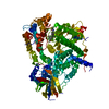
Spodoptera frugiperda (fall armyworm)
References: UniProt: P48736, phosphatidylinositol-4,5-bisphosphate 3-kinase
#2: Chemical ChemComp-FAZ / 8-(acetylamino)-N-(2-chlorophenyl)-N-methyl-4,5-dihydrothieno[3,2-d][1]benzoxepine-2-carboxamide


Mass: 426.916 Da / Num. of mol.: 1 / Source method: obtained synthetically / Formula: C22H19ClN2O3S

-
Experimental details

-
Experiment

ExperimentMethod: X-RAY DIFFRACTION / Number of used crystals: 1

-
Sample preparation

CrystalDensity Matthews: 2.27 Å3/Da / Density % sol: 45.9 %
Crystal growTemperature: 289 K / Method: vapor diffusion, hanging drop / pH: 8.5
Details: PEG 3350, 0.2M NH4SO4, 0.1M Tris-HCl 8.5, vapor diffusion, hanging drop, temperature 289K

-
Data collection

Diffraction
IDCrystal-ID
11
21
Diffraction source
SourceSiteBeamlineIDWavelength (Å)
SYNCHROTRONALS 5.0.211
21
Detector
TypeIDDetectorDate
MAR1CCDMay 17, 2007
ADSC2CCDNov 6, 2007
Radiation
IDProtocolScattering typeWavelength-ID
1SINGLEx-ray1
2SINGLEx-ray1
Radiation wavelengthWavelength: 1 Å / Relative weight: 1
ReflectionResolution: 2.5→50 Å / Num. obs: 34114 / % possible obs: 98.6 % / Redundancy: 2.8 % / Rmerge(I) obs: 0.042 / Net I/σ(I): 15.7
Reflection shell
Resolution (Å)Redundancy (%)Rmerge(I) obsDiffraction-ID% possible all
2.5-2.592.40.5491,294.6
2.59-2.692.70.4681,298.6
2.69-2.822.90.3461,299.2
2.82-2.962.90.2281,299.2
2.96-3.152.90.1471,299.3
3.15-3.392.90.0871,299.3
3.39-3.732.90.0541,299.4
3.73-4.272.90.0381,299.6
4.27-5.382.80.0281,299.6
5.38-502.70.0231,297.1

-
Processing

Software
NameVersionClassificationNB
PHENIXdev_806refinement
SCALEPACKdata scaling
REFMACrefinement
PDB_EXTRACT3.1data extraction
DENZOdata reduction
RefinementMethod to determine structure: MOLECULAR REPLACEMENT
Starting model: 3e8x
Resolution: 2.9→19.927 Å / Cor.coef. Fo:Fc: 0.948 / Cor.coef. Fo:Fc free: 0.923 / Occupancy max: 1 / Occupancy min: 1 / SU ML: 0.62 / SU R Cruickshank DPI: 0.6749 / σ(F): 1.34 / Phase error: 27.89 / Stereochemistry target values: ML / Details: HYDROGENS HAVE BEEN ADDED IN THE RIDING POSITIONS
RfactorNum. reflection% reflection
Rfree0.2531 1179 5.15 %
Rwork0.207 --
obs0.2094 22879 99.7 %
Solvent computationShrinkage radii: 0.9 Å / VDW probe radii: 1.11 Å / Solvent model: FLAT BULK SOLVENT MODEL / Bsol: 60.774 Å2 / ksol: 0.272 e/Å3
Displacement parameters
Baniso -1Baniso -2Baniso -3
1-3.6067 Å20 Å27.4414 Å2
2--2.6581 Å2-0 Å2
3----6.2648 Å2
Refinement stepCycle: LAST / Resolution: 2.9→19.927 Å
ProteinNucleic acidLigandSolventTotal
Num. atoms6775 0 29 0 6804
Refine LS restraints
Refine-IDTypeDev idealNumber
X-RAY DIFFRACTIONf_bond_d0.0116952
X-RAY DIFFRACTIONf_angle_d1.5629408
X-RAY DIFFRACTIONf_dihedral_angle_d16.9552581
X-RAY DIFFRACTIONf_chiral_restr0.1161055
X-RAY DIFFRACTIONf_plane_restr0.011196
LS refinement shell
Resolution (Å)Rfactor RfreeNum. reflection RfreeRfactor RworkNum. reflection RworkRefine-ID% reflection obs (%)
2.9-3.03160.34391590.28472678X-RAY DIFFRACTION100
3.0316-3.19080.31431570.27582702X-RAY DIFFRACTION100
3.1908-3.38980.34131200.26022720X-RAY DIFFRACTION100
3.3898-3.650.3071550.23842690X-RAY DIFFRACTION100
3.65-4.01460.23731440.21332714X-RAY DIFFRACTION100
4.0146-4.58940.19461560.17832693X-RAY DIFFRACTION100
4.5894-5.75890.26971470.18982743X-RAY DIFFRACTION100
5.7589-19.92710.23141410.19332760X-RAY DIFFRACTION99
Refinement TLS params.Method: refined / Origin x: 32.8851 Å / Origin y: 47.0431 Å / Origin z: 26.4657 Å
111213212223313233
T-0.0941 Å20.0309 Å20.0152 Å2--0.3167 Å20.0125 Å2---0.2466 Å2
L4.1281 °2-0.6136 °20.583 °2-1.1076 °2-0.0931 °2--2.045 °2
S-0.1221 Å °-0.353 Å °0.0495 Å °0.2687 Å °0.1715 Å °0.2089 Å °-0.2518 Å °-0.1837 Å °-0.0494 Å °

+
About Yorodumi

-
News

-
Feb 9, 2022. New format data for meta-information of EMDB entries

New format data for meta-information of EMDB entries

  • Version 3 of the EMDB header file is now the official format.
  • The previous official version 1.9 will be removed from the archive.

Related info.:EMDB header

External links:wwPDB to switch to version 3 of the EMDB data model

-
Aug 12, 2020. Covid-19 info

Covid-19 info

URL: https://pdbj.org/emnavi/covid19.php

New page: Covid-19 featured information page in EM Navigator.

Related info.:Covid-19 info / Mar 5, 2020. Novel coronavirus structure data

+
Mar 5, 2020. Novel coronavirus structure data

Novel coronavirus structure data

Related info.:Yorodumi Speices / Aug 12, 2020. Covid-19 info

External links:COVID-19 featured content - PDBj / Molecule of the Month (242):Coronavirus Proteases

+
Jan 31, 2019. EMDB accession codes are about to change! (news from PDBe EMDB page)

EMDB accession codes are about to change! (news from PDBe EMDB page)

  • The allocation of 4 digits for EMDB accession codes will soon come to an end. Whilst these codes will remain in use, new EMDB accession codes will include an additional digit and will expand incrementally as the available range of codes is exhausted. The current 4-digit format prefixed with “EMD-” (i.e. EMD-XXXX) will advance to a 5-digit format (i.e. EMD-XXXXX), and so on. It is currently estimated that the 4-digit codes will be depleted around Spring 2019, at which point the 5-digit format will come into force.
  • The EM Navigator/Yorodumi systems omit the EMD- prefix.

Related info.:Q: What is EMD? / ID/Accession-code notation in Yorodumi/EM Navigator

External links:EMDB Accession Codes are Changing Soon! / Contact to PDBj

+
Jul 12, 2017. Major update of PDB

Major update of PDB

  • wwPDB released updated PDB data conforming to the new PDBx/mmCIF dictionary.
  • This is a major update changing the version number from 4 to 5, and with Remediation, in which all the entries are updated.
  • In this update, many items about electron microscopy experimental information are reorganized (e.g. em_software).
  • Now, EM Navigator and Yorodumi are based on the updated data.

External links:wwPDB Remediation / Enriched Model Files Conforming to OneDep Data Standards Now Available in the PDB FTP Archive

-
Yorodumi

Thousand views of thousand structures

  • Yorodumi is a browser for structure data from EMDB, PDB, SASBDB, etc.
  • This page is also the successor to EM Navigator detail page, and also detail information page/front-end page for Omokage search.
  • The word "yorodu" (or yorozu) is an old Japanese word meaning "ten thousand". "mi" (miru) is to see.

Related info.:EMDB / PDB / SASBDB / Comparison of 3 databanks / Yorodumi Search / Aug 31, 2016. New EM Navigator & Yorodumi / Yorodumi Papers / Jmol/JSmol / Function and homology information / Changes in new EM Navigator and Yorodumi

Read more