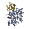
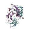
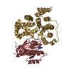
- PDB-3bxj: Crystal Structure of the C2-GAP Fragment of synGAP -
+
Open data
ID or keywords:
Loading...
-
Basic information
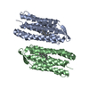
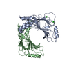
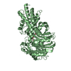
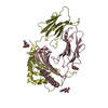
Entry
Database: PDB / ID: 3bxj
Title
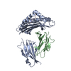
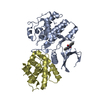
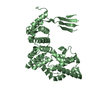
Crystal Structure of the C2-GAP Fragment of synGAP
Components
Ras GTPase-activating protein SynGAP
Keywords
SIGNALING PROTEIN / GTPase activating protein / GTPase activation / Membrane / Phosphoprotein / SH3-binding
Function / homology
Function and homology information
Regulation of RAS by GAPs / negative regulation of axonogenesis / regulation of intracellular signal transduction / maintenance of postsynaptic specialization structure / pattern specification process / negative regulation of Ras protein signal transduction / regulation of synapse structure or activity / receptor clustering / dendrite development / regulation of MAPK cascade ...Regulation of RAS by GAPs / negative regulation of axonogenesis / regulation of intracellular signal transduction / maintenance of postsynaptic specialization structure / pattern specification process / negative regulation of Ras protein signal transduction / regulation of synapse structure or activity / receptor clustering / dendrite development / regulation of MAPK cascade / postsynaptic density, intracellular component / GTPase activator activity / dendritic shaft / SH3 domain binding / visual learning / regulation of synaptic plasticity / regulation of long-term neuronal synaptic plasticity / modulation of chemical synaptic transmission / negative regulation of neuron apoptotic process / Ras protein signal transduction / postsynaptic density / synapse / protein kinase binding / glutamatergic synapse / membrane / plasma membrane Similarity search - Function
In the structure databanks used in Yorodumi, some data are registered as the other names, "COVID-19 virus" and "2019-nCoV". Here are the details of the virus and the list of structure data.
Jan 31, 2019. EMDB accession codes are about to change! (news from PDBe EMDB page)
EMDB accession codes are about to change! (news from PDBe EMDB page)
The allocation of 4 digits for EMDB accession codes will soon come to an end. Whilst these codes will remain in use, new EMDB accession codes will include an additional digit and will expand incrementally as the available range of codes is exhausted. The current 4-digit format prefixed with “EMD-” (i.e. EMD-XXXX) will advance to a 5-digit format (i.e. EMD-XXXXX), and so on. It is currently estimated that the 4-digit codes will be depleted around Spring 2019, at which point the 5-digit format will come into force.
The EM Navigator/Yorodumi systems omit the EMD- prefix.
Related info.:Q: What is EMD? / ID/Accession-code notation in Yorodumi/EM Navigator
Yorodumi is a browser for structure data from EMDB, PDB, SASBDB, etc.
This page is also the successor to EM Navigator detail page, and also detail information page/front-end page for Omokage search.
The word "yorodu" (or yorozu) is an old Japanese word meaning "ten thousand". "mi" (miru) is to see.
Related info.:EMDB / PDB / SASBDB / Comparison of 3 databanks / Yorodumi Search / Aug 31, 2016. New EM Navigator & Yorodumi / Yorodumi Papers / Jmol/JSmol / Function and homology information / Changes in new EM Navigator and Yorodumi
 Movie
Movie Controller


 Open data
 Basic information
 Components
 Keywords
 Function and homology information
 X-RAY DIFFRACTION /
  Authors
 Citation
 Structure visualization
 Downloads & links
 Other downloads
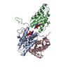
 PDBj
PDBj


 Assembly
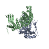


 Sample preparation
 / Beamline: ID14-2 / Wavelength: 0.933 Å
 Processing