 Movie
Movie Controller

[English] 日本語
 Yorodumi
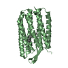
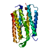
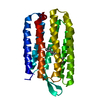
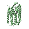
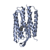
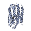
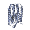
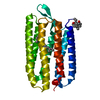
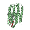
- PDB-5vn9: Structure of bacteriorhodopsin from crystals grown at 4 deg C usi... -
+ Open data

- Basic information
| Entry | Database: PDB / ID: 5vn9 | ||||||
|---|---|---|---|---|---|---|---|
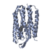
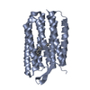
| Title | Structure of bacteriorhodopsin from crystals grown at 4 deg C using GlyNCOC15+4 as an LCP host lipid | ||||||
 Components Components | Bacteriorhodopsin | ||||||
 Keywords Keywords | MEMBRANE PROTEIN / 7TM / retinal protein | ||||||
| Function / homology |  Function and homology information Function and homology informationlight-driven active monoatomic ion transmembrane transporter activity / photoreceptor activity / phototransduction / monoatomic ion channel activity / proton transmembrane transport / plasma membrane Similarity search - Function | ||||||
| Biological species |  Halobacterium salinarum (Halophile) Halobacterium salinarum (Halophile) | ||||||
| Method |  X-RAY DIFFRACTION / SYNCHROTRON / MOLECULAR REPLACEMENT / Resolution: 2.594 Å X-RAY DIFFRACTION / SYNCHROTRON / MOLECULAR REPLACEMENT / Resolution: 2.594 Å | ||||||
 Authors Authors | Ishchenko, A. / Peng, L. / Zinovev, E. / Vlasov, A. / Lee, S.C. / Kuklin, A. / Mishin, A. / Borshchevskiy, V. / Zhang, Q. / Cherezov, V. | ||||||
 Citation Citation | Journal: Cryst Growth Des / Year: 2017 Title: Chemically Stable Lipids for Membrane Protein Crystallization. Authors: Ishchenko, A. / Peng, L. / Zinovev, E. / Vlasov, A. / Lee, S.C. / Kuklin, A. / Mishin, A. / Borshchevskiy, V. / Zhang, Q. / Cherezov, V. | ||||||
| History |
|
- Structure visualization
| Structure viewer | Molecule: MolmilJmol/JSmol |
|---|
- Downloads & links
-Download
| PDBx/mmCIF format | 5vn9.cif.gz | 171.9 KB | Display | PDBx/mmCIF format |
|---|---|---|---|---|
| PDB format | pdb5vn9.ent.gz | 136.8 KB | Display | PDB format |
| PDBx/mmJSON format | 5vn9.json.gz | Tree view | PDBx/mmJSON format | |
| Others |  Other downloads Other downloads |
-Validation report
| Arichive directory | https://data.pdbj.org/pub/pdb/validation_reports/vn/5vn9ftp://data.pdbj.org/pub/pdb/validation_reports/vn/5vn9 | HTTPS FTP |
|---|
-Related structure data
| Related structure data |  5vn7C 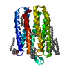 1xjiS C: citing same article ( S: Starting model for refinement |
|---|---|
| Similar structure data |


-Links
 PDBj
PDBj

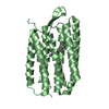
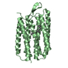
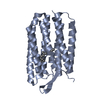
- Assembly
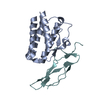
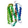
| Deposited unit | 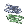
| ||||||||
|---|---|---|---|---|---|---|---|---|---|
| 1 | 
| ||||||||
| 2 | 
| ||||||||
| Unit cell |
|
-Components
| #1: Protein | Mass: 28537.510 Da / Num. of mol.: 2 Source method: isolated from a genetically manipulated source Source: (gene. exp.) Halobacterium salinarum (strain ATCC 700922 / JCM 11081 / NRC-1) (Halophile)Strain: ATCC 700922 / JCM 11081 / NRC-1 / Gene: bop, VNG_1467G / Production host: Halobacterium salinarum (Halophile) / References: UniProt: P02945#2: Water | ChemComp-HOH / |  Mass: 18.015 Da / Num. of mol.: 17 / Source method: isolated from a natural source / Formula: H2O Mass: 18.015 Da / Num. of mol.: 17 / Source method: isolated from a natural source / Formula: H2O |
|---|
-Experimental details
-Experiment
| Experiment | Method: X-RAY DIFFRACTION / Number of used crystals: 1 |
|---|
- Sample preparation
| Crystal | Density Matthews: 2.46 Å3/Da / Density % sol: 50.04 % |
|---|---|
| Crystal grow | Temperature: 277 K / Method: lipidic cubic phase / pH: 5.6 Details: 1.9-3.0 M sodium/potassium phosphate buffer (pH5.6), 3.5% v/v methylpentanediol and 0.5% w/v OG |
-Data collection
| Diffraction | Mean temperature: 100 K |
|---|---|
| Diffraction source | Source: SYNCHROTRON / Site: APS  / Beamline: 23-ID-D / Wavelength: 1.033 Å / Beamline: 23-ID-D / Wavelength: 1.033 Å |
| Detector | Type: DECTRIS PILATUS3 S 6M / Detector: PIXEL / Date: Apr 1, 2015 |
| Radiation | Protocol: SINGLE WAVELENGTH / Monochromatic (M) / Laue (L): M / Scattering type: x-ray |
| Radiation wavelength | Wavelength: 1.033 Å / Relative weight: 1 |
| Reflection | Resolution: 2.59→28.342 Å / Num. obs: 15034 / % possible obs: 90 % / Redundancy: 2.7 % / Biso Wilson estimate: 46.5 Å2 / CC1/2: 0.987 / Rmerge(I) obs: 0.143 / Net I/σ(I): 5.3 |
| Reflection shell | Resolution: 2.59→2.69 Å / Redundancy: 1.7 % / Rmerge(I) obs: 0.317 / Mean I/σ(I) obs: 1.7 / Num. unique obs: 1396 / CC1/2: 0.76 / % possible all: 50 |
- Processing
| Software |
| ||||||||||||||||||||||||||||||||||||||||||
|---|---|---|---|---|---|---|---|---|---|---|---|---|---|---|---|---|---|---|---|---|---|---|---|---|---|---|---|---|---|---|---|---|---|---|---|---|---|---|---|---|---|---|---|
| Refinement | Method to determine structure: MOLECULAR REPLACEMENT Starting model: 1XJI Resolution: 2.594→28.342 Å / Cross valid method: FREE R-VALUE / σ(F): 1.97 / Phase error: 28.87 / Stereochemistry target values: TWIN_LSQ_F
| ||||||||||||||||||||||||||||||||||||||||||
| Solvent computation | Shrinkage radii: 0.9 Å / VDW probe radii: 1.11 Å / Solvent model: FLAT BULK SOLVENT MODEL | ||||||||||||||||||||||||||||||||||||||||||
| Refinement step | Cycle: LAST / Resolution: 2.594→28.342 Å
| ||||||||||||||||||||||||||||||||||||||||||
| Refine LS restraints |
| ||||||||||||||||||||||||||||||||||||||||||
| LS refinement shell |
|
