[English] 日本語
Yorodumi
- PDB-6axz: Segment from bank vole prion protein 168-176 QYNNQNNFV -

+
Open data


ID or keywords:

Loading...

-
Basic information

Entry
Database: PDB / ID: 6axz
TitleSegment from bank vole prion protein 168-176 QYNNQNNFV
ComponentsMajor prion protein
KeywordsPROTEIN FIBRIL / polar clasp / amyloid fibril / prion
Function / homology
Function and homology information


side of membrane / protein homooligomerization / Golgi apparatus / metal ion binding / plasma membrane
Similarity search - Function
Prion protein signature 1. / Prion protein signature 2. / Major prion protein N-terminal domain / Major prion protein bPrPp - N terminal / Prion protein / Major prion protein / Prion/Doppel protein, beta-ribbon domain / Prion/Doppel beta-ribbon domain superfamily / Prion/Doppel alpha-helical domain
Similarity search - Domain/homology
Biological speciesMyodes glareolus (Bank vole)
MethodELECTRON CRYSTALLOGRAPHY / electron crystallography / AB INITIO PHASING / cryo EM / Resolution: 0.75 Å
Model detailspolar clasp, MicroED, Glutamine ladder, Asparagine ladder
AuthorsGlynn, C. / Rodriguez, J.A. / Boyer, D.R. / Gallagher-Jones, M.
Citation
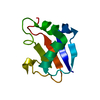
Journal: Nat Struct Mol Biol / Year: 2018
Title: Sub-ångström cryo-EM structure of a prion protofibril reveals a polar clasp.
Authors: Marcus Gallagher-Jones / Calina Glynn / David R Boyer / Michael W Martynowycz / Evelyn Hernandez / Jennifer Miao / Chih-Te Zee / Irina V Novikova / Lukasz Goldschmidt / Heather T McFarlane / ...Authors: Marcus Gallagher-Jones / Calina Glynn / David R Boyer / Michael W Martynowycz / Evelyn Hernandez / Jennifer Miao / Chih-Te Zee / Irina V Novikova / Lukasz Goldschmidt / Heather T McFarlane / Gustavo F Helguera / James E Evans / Michael R Sawaya / Duilio Cascio / David S Eisenberg / Tamir Gonen / Jose A Rodriguez /
Abstract: The atomic structure of the infectious, protease-resistant, β-sheet-rich and fibrillar mammalian prion remains unknown. Through the cryo-EM method MicroED, we reveal the sub-ångström-resolution ...The atomic structure of the infectious, protease-resistant, β-sheet-rich and fibrillar mammalian prion remains unknown. Through the cryo-EM method MicroED, we reveal the sub-ångström-resolution structure of a protofibril formed by a wild-type segment from the β2-α2 loop of the bank vole prion protein. The structure of this protofibril reveals a stabilizing network of hydrogen bonds that link polar zippers within a sheet, producing motifs we have named 'polar clasps'.
History
DepositionSep 7, 2017Deposition site: RCSB / Processing site: RCSB
Revision 1.0Jan 17, 2018Provider: repository / Type: Initial release
Revision 1.1Jan 31, 2018Group: Database references / Category: citation / citation_author
Item: _citation.journal_abbrev / _citation.pdbx_database_id_PubMed ..._citation.journal_abbrev / _citation.pdbx_database_id_PubMed / _citation.title / _citation_author.name
Revision 1.2Feb 21, 2018Group: Database references / Category: citation
Item: _citation.journal_volume / _citation.page_first / _citation.page_last
Revision 1.3Mar 13, 2024Group: Data collection / Database references
Category: chem_comp_atom / chem_comp_bond ...chem_comp_atom / chem_comp_bond / database_2 / diffrn_radiation_wavelength
Item: _database_2.pdbx_DOI / _database_2.pdbx_database_accession / _diffrn_radiation_wavelength.wavelength

-
Structure visualization

Movie
  • Biological unit as author_defined_assembly
  • Imaged by Jmol
  • Download
  • Deposited structure unit
  • Imaged by Jmol
  • Download
  • Superimposition on EM map
  • EMDB-7017
  • Imaged by UCSF Chimera
  • Download
Movie viewer
Structure viewerMolecule:
MolmilJmol/JSmol

Downloads & links

-
Assembly

Deposited unit
A: Major prion protein


Theoretical massNumber of molelcules
Total (without water)1,1401
Polymers1,1401
Non-polymers00
Water362
1
A: Major prion protein

A: Major prion protein

A: Major prion protein

A: Major prion protein

A: Major prion protein

A: Major prion protein

A: Major prion protein

A: Major prion protein

A: Major prion protein

A: Major prion protein


Theoretical massNumber of molelcules
Total (without water)11,40210
Polymers11,40210
Non-polymers00
Water18010
TypeNameSymmetry operationNumber
identity operation1_555x,y,z1
crystal symmetry operation1_655x+1,y,z1
crystal symmetry operation1_755x+2,y,z1
crystal symmetry operation1_855x+3,y,z1
crystal symmetry operation1_955x+4,y,z1
crystal symmetry operation1_565x,y+1,z1
crystal symmetry operation1_665x+1,y+1,z1
crystal symmetry operation1_765x+2,y+1,z1
crystal symmetry operation1_865x+3,y+1,z1
crystal symmetry operation1_965x+4,y+1,z1
Unit cell
Length a, b, c (Å)4.940, 10.340, 31.150
Angle α, β, γ (deg.)94.210, 92.380, 102.200
Int Tables number1
Space group name H-MP1

-
Components

#1: Protein/peptide Major prion protein


Mass: 1140.162 Da / Num. of mol.: 1 / Fragment: UNP residues 168-176 / Source method: obtained synthetically / Source: (synth.) Myodes glareolus (Bank vole) / References: UniProt: Q8VHV5
#2: Water ChemComp-HOH / water / Water


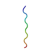
Mass: 18.015 Da / Num. of mol.: 2 / Source method: isolated from a natural source / Formula: H2O

-
Experimental details

-
Experiment

ExperimentMethod: ELECTRON CRYSTALLOGRAPHY / Number of used crystals: 1
EM experimentAggregation state: 3D ARRAY / 3D reconstruction method: electron crystallography

-
Sample preparation

ComponentName: bank vole prion 168-176 / Type: COMPLEX / Entity ID: #1 / Source: NATURAL
Molecular weightValue: 0.0046 MDa / Experimental value: YES
Source (natural)Organism: Myodes glareolus (Bank vole)
Buffer solutionpH: 6
SpecimenEmbedding applied: NO / Shadowing applied: NO / Staining applied: NO / Vitrification applied: YES
VitrificationCryogen name: ETHANE
Crystal growTemperature: 298 K / Method: batch mode / pH: 6 / Details: 10% MPD, 0.1 M MES, pH 6.0

-
Data collection

Experimental equipment
Model: Tecnai F20 / Image courtesy: FEI Company
MicroscopyModel: FEI TECNAI F20
Electron gunElectron source: FIELD EMISSION GUN / Accelerating voltage: 200 kV / Illumination mode: FLOOD BEAM
Electron lensMode: DIFFRACTION
Image recordingElectron dose: 0.025 e/Å2 / Film or detector model: TVIPS TEMCAM-F416 (4k x 4k)
EM diffractionCamera length: 950 mm
EM diffraction shellResolution: 0.75→0.77 Å / Fourier space coverage: 96.2 % / Multiplicity: 4.4 / Num. of structure factors: 532 / Phase residual: 0.01 °
EM diffraction statsFourier space coverage: 97.1 % / High resolution: 0.75 Å / Num. of intensities measured: 43252 / Num. of structure factors: 7474 / Phase error: 0.01 ° / Phase residual: 0.01 ° / Phase error rejection criteria: 0 / Rmerge: 23.2 / Rsym: 23.2
DiffractionMean temperature: 100 K
RadiationProtocol: SINGLE WAVELENGTH / Monochromatic (M) / Laue (L): M / Scattering type: electron
Radiation wavelengthRelative weight: 1
ReflectionResolution: 0.75→10.338 Å / Num. obs: 7474 / % possible obs: 97.1 % / Observed criterion σ(I): -3 / Redundancy: 5.787 % / Biso Wilson estimate: 4.17 Å2 / CC1/2: 0.982 / Rmerge(I) obs: 0.232 / Rrim(I) all: 0.25 / Χ2: 0.872 / Net I/σ(I): 4.57 / Num. measured all: 43252 / Scaling rejects: 7
Reflection shell

Diffraction-ID: 1

Resolution (Å)Redundancy (%)Rmerge(I) obsMean I/σ(I) obsNum. measured obsNum. possibleNum. unique obsCC1/2Rrim(I) all% possible all
0.75-0.774.4340.6381.7723595535320.2090.72996.2
0.77-0.794.9750.5812.3327715825570.3830.65595.7
0.79-0.815.2250.5432.5727385495240.4830.60695.4
0.81-0.845.0460.5142.5725185204990.510.57996
0.84-0.875.1340.4792.8424444894760.6110.53597.3
0.87-0.95.4120.4263.2625224874660.7750.47195.7
0.9-0.935.6850.3773.8326324794630.8330.41496.7
0.93-0.976.1630.3634.4228354754600.8660.39396.8
0.97-1.016.2450.3124.9227044434330.9170.33697.7
1.01-1.065.8190.3364.922813983920.8910.36698.5
1.06-1.126.1140.2855.4923603883860.9460.30999.5
1.12-1.196.8260.2746.4525943853800.9390.29498.7
1.19-1.276.7790.266.5325493783760.9490.2899.5
1.27-1.376.0030.2576.417472992910.930.2897.3
1.37-1.56.630.2437.1220223113050.950.26198.1
1.5-1.686.8760.2167.818912872750.960.23195.8
1.68-1.946.0540.2017.5112412112050.9670.21897.2
1.94-2.376.7730.2098.2214902222200.9650.22599.1
2.37-3.356.570.1688.49921551510.9820.1897.4
3.35-10.3386.7710.2058.656288830.9680.21894.3

-
Processing

Software
NameVersionClassification
PHENIXrefinement
XSCALEdata scaling
PDB_EXTRACT3.22data extraction
EM softwareName: Coot / Category: model refinement
EM 3D crystal entity∠α: 94.21 ° / ∠β: 92.38 ° / ∠γ: 102.2 ° / A: 4.94 Å / B: 10.34 Å / C: 31.15 Å / Space group name: P1 / Space group num: 1
CTF correctionType: NONE
3D reconstructionResolution method: DIFFRACTION PATTERN/LAYERLINES / Symmetry type: 3D CRYSTAL
Atomic model buildingB value: 6.068 / Protocol: AB INITIO MODEL / Space: RECIPROCAL / Target criteria: maximum likelihood
RefinementMethod to determine structure: AB INITIO PHASING / Resolution: 0.75→10.338 Å / SU ML: 0.15 / Cross valid method: FREE R-VALUE / σ(F): 1.95 / Phase error: 36.9 / Stereochemistry target values: ML
RfactorNum. reflection% reflectionSelection details
Rfree0.246 673 9.01 %0
Rwork0.2422 6800 --
obs0.2426 7473 96.96 %-
Solvent computationShrinkage radii: 0.9 Å / VDW probe radii: 1.11 Å / Solvent model: FLAT BULK SOLVENT MODEL
Displacement parametersBiso max: 17.71 Å2 / Biso mean: 6.0686 Å2 / Biso min: 1.29 Å2
Refine LS restraints
Refine-IDTypeDev idealNumber
ELECTRON CRYSTALLOGRAPHYf_bond_d0.01482
ELECTRON CRYSTALLOGRAPHYf_angle_d1.014111
ELECTRON CRYSTALLOGRAPHYf_chiral_restr0.10410
ELECTRON CRYSTALLOGRAPHYf_plane_restr0.00517
ELECTRON CRYSTALLOGRAPHYf_dihedral_angle_d18.83829
LS refinement shell

Refine-ID: ELECTRON CRYSTALLOGRAPHY / Rfactor Rfree error: 0 / Total num. of bins used: 5

Resolution (Å)Rfactor RfreeNum. reflection RfreeRfactor RworkNum. reflection RworkNum. reflection all% reflection obs (%)
0.7495-0.80740.39071340.36831358149295
0.8074-0.88860.36621300.34421313144397
0.8886-1.01710.28591370.28591383152097
1.0171-1.2810.2621370.24361391152899
1.281-10.33870.18511350.18751355149097

+
About Yorodumi

-
News

-
Feb 9, 2022. New format data for meta-information of EMDB entries

New format data for meta-information of EMDB entries

  • Version 3 of the EMDB header file is now the official format.
  • The previous official version 1.9 will be removed from the archive.

Related info.:EMDB header

External links:wwPDB to switch to version 3 of the EMDB data model

-
Aug 12, 2020. Covid-19 info

Covid-19 info

URL: https://pdbj.org/emnavi/covid19.php

New page: Covid-19 featured information page in EM Navigator.

Related info.:Covid-19 info / Mar 5, 2020. Novel coronavirus structure data

+
Mar 5, 2020. Novel coronavirus structure data

Novel coronavirus structure data

Related info.:Yorodumi Speices / Aug 12, 2020. Covid-19 info

External links:COVID-19 featured content - PDBj / Molecule of the Month (242):Coronavirus Proteases

+
Jan 31, 2019. EMDB accession codes are about to change! (news from PDBe EMDB page)

EMDB accession codes are about to change! (news from PDBe EMDB page)

  • The allocation of 4 digits for EMDB accession codes will soon come to an end. Whilst these codes will remain in use, new EMDB accession codes will include an additional digit and will expand incrementally as the available range of codes is exhausted. The current 4-digit format prefixed with “EMD-” (i.e. EMD-XXXX) will advance to a 5-digit format (i.e. EMD-XXXXX), and so on. It is currently estimated that the 4-digit codes will be depleted around Spring 2019, at which point the 5-digit format will come into force.
  • The EM Navigator/Yorodumi systems omit the EMD- prefix.

Related info.:Q: What is EMD? / ID/Accession-code notation in Yorodumi/EM Navigator

External links:EMDB Accession Codes are Changing Soon! / Contact to PDBj

+
Jul 12, 2017. Major update of PDB

Major update of PDB

  • wwPDB released updated PDB data conforming to the new PDBx/mmCIF dictionary.
  • This is a major update changing the version number from 4 to 5, and with Remediation, in which all the entries are updated.
  • In this update, many items about electron microscopy experimental information are reorganized (e.g. em_software).
  • Now, EM Navigator and Yorodumi are based on the updated data.

External links:wwPDB Remediation / Enriched Model Files Conforming to OneDep Data Standards Now Available in the PDB FTP Archive

-
Yorodumi

Thousand views of thousand structures

  • Yorodumi is a browser for structure data from EMDB, PDB, SASBDB, etc.
  • This page is also the successor to EM Navigator detail page, and also detail information page/front-end page for Omokage search.
  • The word "yorodu" (or yorozu) is an old Japanese word meaning "ten thousand". "mi" (miru) is to see.

Related info.:EMDB / PDB / SASBDB / Comparison of 3 databanks / Yorodumi Search / Aug 31, 2016. New EM Navigator & Yorodumi / Yorodumi Papers / Jmol/JSmol / Function and homology information / Changes in new EM Navigator and Yorodumi

Read more