 Movie
Movie Controller


+ Open data

- Basic information
| Entry | Database: EMDB / ID: EMD-7017 | |||||||||
|---|---|---|---|---|---|---|---|---|---|---|
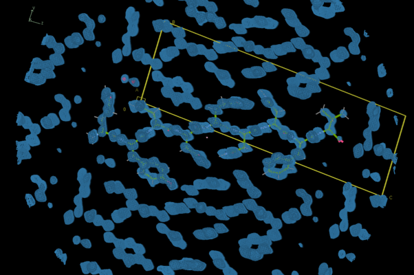
| Title | Segment from bank vole prion protein 168-176 QYNNQNNFV | |||||||||
 Map data Map data | protein segment | |||||||||
 Sample Sample |
| |||||||||
 Keywords Keywords | polar clasp / amyloid fibril / prion / PROTEIN FIBRIL | |||||||||
| Function / homology |  Function and homology information Function and homology informationside of membrane / protein homooligomerization / Golgi apparatus / metal ion binding / plasma membrane Similarity search - Function | |||||||||
| Biological species |  Myodes glareolus (Bank vole) Myodes glareolus (Bank vole) | |||||||||
| Method | electron crystallography / cryo EM | |||||||||
 Authors Authors | Glynn C / Rodriguez JA | |||||||||
 Citation Citation | Journal: Nat Struct Mol Biol / Year: 2018 Title: Sub-ångström cryo-EM structure of a prion protofibril reveals a polar clasp. Authors: Marcus Gallagher-Jones / Calina Glynn / David R Boyer / Michael W Martynowycz / Evelyn Hernandez / Jennifer Miao / Chih-Te Zee / Irina V Novikova / Lukasz Goldschmidt / Heather T McFarlane / ...Authors: Marcus Gallagher-Jones / Calina Glynn / David R Boyer / Michael W Martynowycz / Evelyn Hernandez / Jennifer Miao / Chih-Te Zee / Irina V Novikova / Lukasz Goldschmidt / Heather T McFarlane / Gustavo F Helguera / James E Evans / Michael R Sawaya / Duilio Cascio / David S Eisenberg / Tamir Gonen / Jose A Rodriguez /   Abstract: The atomic structure of the infectious, protease-resistant, β-sheet-rich and fibrillar mammalian prion remains unknown. Through the cryo-EM method MicroED, we reveal the sub-ångström-resolution ...The atomic structure of the infectious, protease-resistant, β-sheet-rich and fibrillar mammalian prion remains unknown. Through the cryo-EM method MicroED, we reveal the sub-ångström-resolution structure of a protofibril formed by a wild-type segment from the β2-α2 loop of the bank vole prion protein. The structure of this protofibril reveals a stabilizing network of hydrogen bonds that link polar zippers within a sheet, producing motifs we have named 'polar clasps'. | |||||||||
| History |
|
- Structure visualization
| Movie |
Movie viewer |
|---|---|
| Structure viewer | EM map: SurfViewMolmilJmol/JSmol |
| Supplemental images |
- Downloads & links
-EMDB archive
| Map data | emd_7017.map.gz | 448.6 KB | EMDB map data format | |
|---|---|---|---|---|
| Header (meta data) | emd-7017-v30.xmlemd-7017.xml | 10.5 KB 10.5 KB | Display Display | EMDB header |
| Images |  emd_7017.png emd_7017.png | 185.4 KB | ||
| Filedesc metadata | emd-7017.cif.gz | 4.3 KB | ||
| Filedesc structureFactors | emd_7017_sf.cif.gz | 123 KB | ||
| Archive directory |  http://ftp.pdbj.org/pub/emdb/structures/EMD-7017ftp://ftp.pdbj.org/pub/emdb/structures/EMD-7017 http://ftp.pdbj.org/pub/emdb/structures/EMD-7017ftp://ftp.pdbj.org/pub/emdb/structures/EMD-7017 | HTTPS FTP |
-Related structure data
| Related structure data |  6axzMC  7287C  6btkC M: atomic model generated by this map C: citing same article ( |
|---|---|
| Similar structure data |
-Links
| EMDB pages | EMDB (EBI/PDBe) / EMDataResource |
|---|---|
| Related items in Molecule of the Month |


-Map
| File | Download / File: emd_7017.map.gz / Format: CCP4 / Size: 484.4 KB / Type: IMAGE STORED AS FLOATING POINT NUMBER (4 BYTES) | ||||||||||||||||||||||||||||||||||||||||||||||||||||||||||||||||||||
|---|---|---|---|---|---|---|---|---|---|---|---|---|---|---|---|---|---|---|---|---|---|---|---|---|---|---|---|---|---|---|---|---|---|---|---|---|---|---|---|---|---|---|---|---|---|---|---|---|---|---|---|---|---|---|---|---|---|---|---|---|---|---|---|---|---|---|---|---|---|
| Annotation | protein segment | ||||||||||||||||||||||||||||||||||||||||||||||||||||||||||||||||||||
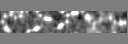
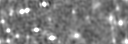
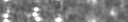
| Projections & slices | Image control
Images are generated by Spider. generated in cubic-lattice coordinate | ||||||||||||||||||||||||||||||||||||||||||||||||||||||||||||||||||||
| Voxel size | X: 0.22455 Å / Y: 0.235 Å / Z: 0.24336 Å | ||||||||||||||||||||||||||||||||||||||||||||||||||||||||||||||||||||
| Density |
| ||||||||||||||||||||||||||||||||||||||||||||||||||||||||||||||||||||
| Symmetry | Space group: 1 | ||||||||||||||||||||||||||||||||||||||||||||||||||||||||||||||||||||
| Details | EMDB XML:
CCP4 map header:
| ||||||||||||||||||||||||||||||||||||||||||||||||||||||||||||||||||||
 Y (Sec.)
 X (Row.)
 Z (Col.)


-Supplemental data
- Sample components
-Entire : bank vole prion 168-176
| Entire | Name: bank vole prion 168-176 |
|---|---|
| Components |
|
-Supramolecule #1: bank vole prion 168-176
| Supramolecule | Name: bank vole prion 168-176 / type: complex / ID: 1 / Parent: 0 / Macromolecule list: #1 |
|---|---|
| Source (natural) | Organism: Myodes glareolus (Bank vole) |
| Molecular weight | Theoretical: 4.6 KDa |
-Macromolecule #1: Major prion protein
| Macromolecule | Name: Major prion protein / type: protein_or_peptide / ID: 1 / Number of copies: 1 / Enantiomer: LEVO |
|---|---|
| Source (natural) | Organism: Myodes glareolus (Bank vole) |
| Molecular weight | Theoretical: 1.140162 KDa |
| Sequence | String: QYNNQNNFV UniProtKB: Major prion protein |
-Macromolecule #2: water
| Macromolecule | Name: water / type: ligand / ID: 2 / Number of copies: 2 / Formula: HOH |
|---|---|
| Molecular weight | Theoretical: 18.015 Da |
| Chemical component information |  ChemComp-HOH: |
-Experimental details
-Structure determination
| Method | cryo EM |
|---|---|
 Processing Processing | electron crystallography |
| Aggregation state | 3D array |
-Sample preparation
| Buffer | pH: 6 |
|---|---|
| Vitrification | Cryogen name: ETHANE |
- Electron microscopy
| Microscope | FEI TECNAI F20 |
|---|---|
| Image recording | Film or detector model: TVIPS TEMCAM-F416 (4k x 4k) / Average electron dose: 0.025 e/Å2 |
| Electron beam | Acceleration voltage: 200 kV / Electron source:  FIELD EMISSION GUN FIELD EMISSION GUN |
| Electron optics | Illumination mode: FLOOD BEAM / Imaging mode: DIFFRACTION / Camera length: 950 mm |
| Experimental equipment |  Model: Tecnai F20 / Image courtesy: FEI Company |
-Image processing
| Final reconstruction | Resolution method: DIFFRACTION PATTERN/LAYERLINES |
|---|---|
| Crystallography statistics | Number intensities measured: 43252 / Number structure factors: 7474 / Fourier space coverage: 97.1 / R sym: 23.2 / R merge: 23.2 / Overall phase error: 0.01 / Overall phase residual: 0.01 / Phase error rejection criteria: 0 / High resolution: 0.75 Å / Shell - Shell ID: 1 / Shell - High resolution: 0.75 Å / Shell - Low resolution: 0.77 Å / Shell - Number structure factors: 532 / Shell - Phase residual: 0.01 / Shell - Fourier space coverage: 96.2 / Shell - Multiplicity: 4.4 |
-Atomic model buiding 1
| Refinement | Space: RECIPROCAL / Protocol: AB INITIO MODEL / Overall B value: 6.068 / Target criteria: maximum likelihood |
|---|---|
| Output model | PDB-6axz: |
