 Movie
Movie Controller

[English] 日本語
 Yorodumi
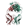
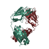
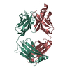
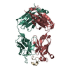
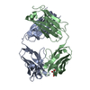
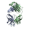
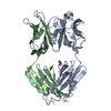
- PDB-6avn: Crystal structure of unbound anti-HIV antibody Fab PGV19 at 2.5 A -
+ Open data

- Basic information
| Entry | Database: PDB / ID: 6avn | ||||||
|---|---|---|---|---|---|---|---|
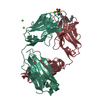
| Title | Crystal structure of unbound anti-HIV antibody Fab PGV19 at 2.5 A | ||||||
 Components Components |
| ||||||
 Keywords Keywords | IMMUNE SYSTEM / VRC01-class lambda antibody / CD4 binding site epitope / anti-HIV neutralizing antibody | ||||||
| Function / homology | Immunoglobulins / Immunoglobulin-like / Sandwich / Mainly Beta Function and homology information Function and homology information | ||||||
| Biological species |  Homo sapiens (human) Homo sapiens (human) | ||||||
| Method |  X-RAY DIFFRACTION / SYNCHROTRON / MOLECULAR REPLACEMENT / Resolution: 2.5 Å X-RAY DIFFRACTION / SYNCHROTRON / MOLECULAR REPLACEMENT / Resolution: 2.5 Å | ||||||
 Authors Authors | Sarkar, A. / Wilson, I.A. | ||||||
 Citation Citation | Journal: Nat Commun / Year: 2018 Title: Structure of a cleavage-independent HIV Env recapitulates the glycoprotein architecture of the native cleaved trimer. Authors: Sarkar, A. / Bale, S. / Behrens, A.J. / Kumar, S. / Sharma, S.K. / de Val, N. / Pallesen, J. / Irimia, A. / Diwanji, D.C. / Stanfield, R.L. / Ward, A.B. / Crispin, M. / Wyatt, R.T. / Wilson, I.A. | ||||||
| History |
|
- Structure visualization
| Structure viewer | Molecule: MolmilJmol/JSmol |
|---|
- Downloads & links
-Download
| PDBx/mmCIF format | 6avn.cif.gz | 179.8 KB | Display | PDBx/mmCIF format |
|---|---|---|---|---|
| PDB format | pdb6avn.ent.gz | 141.4 KB | Display | PDB format |
| PDBx/mmJSON format | 6avn.json.gz | Tree view | PDBx/mmJSON format | |
| Others |  Other downloads Other downloads |
-Validation report
| Arichive directory | https://data.pdbj.org/pub/pdb/validation_reports/av/6avnftp://data.pdbj.org/pub/pdb/validation_reports/av/6avn | HTTPS FTP |
|---|
-Related structure data
| Related structure data |  6b0nC 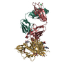 4lsuS S: Starting model for refinement C: citing same article ( |
|---|---|
| Similar structure data |


-Links
 PDBj
PDBj

- Assembly
| Deposited unit | 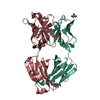
| ||||||||
|---|---|---|---|---|---|---|---|---|---|
| 1 |
| ||||||||
| Unit cell |
|
-Components
| #1: Antibody | Mass: 24246.449 Da / Num. of mol.: 1 Source method: isolated from a genetically manipulated source Source: (gene. exp.) Homo sapiens (human) / Plasmid: phCMV3 / Cell line (production host): HEK 293 Freestyle / Production host: Homo sapiens (human) | ||||||
|---|---|---|---|---|---|---|---|
| #2: Antibody | Mass: 22219.646 Da / Num. of mol.: 1 Source method: isolated from a genetically manipulated source Source: (gene. exp.) Homo sapiens (human) / Plasmid: phCMV3 / Cell line (production host): HEK 293 Freestyle / Production host: Homo sapiens (human) | ||||||
| #3: Chemical | ChemComp-GOL /   Mass: 92.094 Da / Num. of mol.: 4 / Source method: obtained synthetically / Formula: C3H8O3 Mass: 92.094 Da / Num. of mol.: 4 / Source method: obtained synthetically / Formula: C3H8O3#4: Chemical | ChemComp-1PE / |   Mass: 238.278 Da / Num. of mol.: 1 / Source method: obtained synthetically / Formula: C10H22O6 / Comment: precipitant*YM Mass: 238.278 Da / Num. of mol.: 1 / Source method: obtained synthetically / Formula: C10H22O6 / Comment: precipitant*YM#5: Water | ChemComp-HOH / |  Mass: 18.015 Da / Num. of mol.: 153 / Source method: isolated from a natural source / Formula: H2O Mass: 18.015 Da / Num. of mol.: 153 / Source method: isolated from a natural source / Formula: H2OHas protein modification | Y | |
-Experimental details
-Experiment
| Experiment | Method: X-RAY DIFFRACTION / Number of used crystals: 1 |
|---|
- Sample preparation
| Crystal | Density Matthews: 2.53 Å3/Da / Density % sol: 51.42 % |
|---|---|
| Crystal grow | Temperature: 293.15 K / Method: vapor diffusion, sitting drop Details: 10% (v/v) glycerol, 0.1 M ammonium sulfate, 0.1 M HEPES (pH 7.5), 5% (w/v) PEG 3000 and 30% (v/v) PEG 400 |
-Data collection
| Diffraction | Mean temperature: 293 K |
|---|---|
| Diffraction source | Source: SYNCHROTRON / Site: APS  / Beamline: 23-ID-B / Wavelength: 1 Å / Beamline: 23-ID-B / Wavelength: 1 Å |
| Detector | Type: DECTRIS PILATUS3 6M / Detector: PIXEL / Date: Aug 9, 2014 |
| Radiation | Protocol: SINGLE WAVELENGTH / Monochromatic (M) / Laue (L): M / Scattering type: x-ray |
| Radiation wavelength | Wavelength: 1 Å / Relative weight: 1 |
| Reflection | Resolution: 2.5→50 Å / Num. obs: 17498 / % possible obs: 100 % / Redundancy: 7.8 % / Net I/σ(I): 13.2 |
| Reflection shell | Resolution: 2.5→2.54 Å / Redundancy: 7.8 % / Num. unique obs: 837 / % possible all: 100 |
- Processing
| Software |
| |||||||||||||||||||||||||||||||||||||||||||||||||
|---|---|---|---|---|---|---|---|---|---|---|---|---|---|---|---|---|---|---|---|---|---|---|---|---|---|---|---|---|---|---|---|---|---|---|---|---|---|---|---|---|---|---|---|---|---|---|---|---|---|---|
| Refinement | Method to determine structure: MOLECULAR REPLACEMENT Starting model: 4LSU Resolution: 2.5→49.344 Å / SU ML: 0.34 / Cross valid method: FREE R-VALUE / σ(F): 1.34 / Phase error: 23.79 / Stereochemistry target values: ML
| |||||||||||||||||||||||||||||||||||||||||||||||||
| Solvent computation | Shrinkage radii: 0.9 Å / VDW probe radii: 1.11 Å / Solvent model: FLAT BULK SOLVENT MODEL | |||||||||||||||||||||||||||||||||||||||||||||||||
| Refinement step | Cycle: LAST / Resolution: 2.5→49.344 Å
| |||||||||||||||||||||||||||||||||||||||||||||||||
| Refine LS restraints |
| |||||||||||||||||||||||||||||||||||||||||||||||||
| LS refinement shell |
| |||||||||||||||||||||||||||||||||||||||||||||||||
| Refinement TLS params. | Method: refined / Origin x: 32.3809 Å / Origin y: -5.1523 Å / Origin z: -15.4183 Å
| |||||||||||||||||||||||||||||||||||||||||||||||||
| Refinement TLS group | Selection details: all |
