 Movie
Movie Controller

[English] 日本語
 Yorodumi
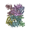
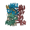
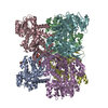
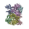
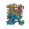
- PDB-5xvi: Crystal Structure of Aspergillus niger Apo- Glutamate Dehydrogenase -
+ Open data

- Basic information
| Entry | Database: PDB / ID: 5xvi | ||||||
|---|---|---|---|---|---|---|---|
| Title | Crystal Structure of Aspergillus niger Apo- Glutamate Dehydrogenase | ||||||
 Components Components | Glutamate dehydrogenase | ||||||
 Keywords Keywords | OXIDOREDUCTASE / Aspergillus / Glutamate / Dehydrogenase / 2-oxoglutarate / Allostery | ||||||
| Function / homology |  Function and homology information Function and homology informationglutamate biosynthetic process / glutamate dehydrogenase (NADP+) activity / nucleotide binding / cytosol Similarity search - Function | ||||||
| Biological species |  | ||||||
| Method |  X-RAY DIFFRACTION / MOLECULAR REPLACEMENT / Resolution: 2.8 Å X-RAY DIFFRACTION / MOLECULAR REPLACEMENT / Resolution: 2.8 Å | ||||||
 Authors Authors | Prakash, P. / Punekar, N.S. / Bhaumik, P. | ||||||
| Funding support |  India, 1items India, 1items
| ||||||
 Citation Citation | Journal: J. Biol. Chem. / Year: 2018 Title: Structural basis for the catalytic mechanism and alpha-ketoglutarate cooperativity of glutamate dehydrogenase. Authors: Prakash, P. / Punekar, N.S. / Bhaumik, P. | ||||||
| History |
|
- Structure visualization
| Structure viewer | Molecule: MolmilJmol/JSmol |
|---|
- Downloads & links
-Download
| PDBx/mmCIF format | 5xvi.cif.gz | 513 KB | Display | PDBx/mmCIF format |
|---|---|---|---|---|
| PDB format | pdb5xvi.ent.gz | 426.1 KB | Display | PDB format |
| PDBx/mmJSON format | 5xvi.json.gz | Tree view | PDBx/mmJSON format | |
| Others |  Other downloads Other downloads |
-Validation report
| Summary document | 5xvi_validation.pdf.gz | 478.8 KB | Display | wwPDB validaton report |
|---|---|---|---|---|
| Full document | 5xvi_full_validation.pdf.gz | 496.3 KB | Display | |
| Data in XML | 5xvi_validation.xml.gz | 94.6 KB | Display | |
| Data in CIF | 5xvi_validation.cif.gz | 133 KB | Display | |
| Arichive directory | https://data.pdbj.org/pub/pdb/validation_reports/xv/5xviftp://data.pdbj.org/pub/pdb/validation_reports/xv/5xvi | HTTPS FTP |
-Related structure data
| Related structure data |  5xvvC 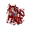 5xvxC 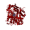 5xw0C 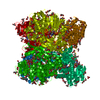 5xwcC 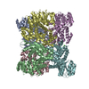 3sboS S: Starting model for refinement C: citing same article ( |
|---|---|
| Similar structure data |


-Links
 PDBj
PDBj- Assembly
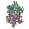
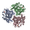
| Deposited unit | 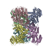
| ||||||||
|---|---|---|---|---|---|---|---|---|---|
| 1 |
| ||||||||
| Unit cell |
|
-Components
| #1: Protein | Mass: 49442.508 Da / Num. of mol.: 6 Source method: isolated from a genetically manipulated source Source: (gene. exp.) Production host: References: UniProt: B6V7E4 #2: Chemical |   Mass: 92.094 Da / Num. of mol.: 2 / Source method: obtained synthetically / Formula: C3H8O3 Mass: 92.094 Da / Num. of mol.: 2 / Source method: obtained synthetically / Formula: C3H8O3#3: Water | ChemComp-HOH / |  Mass: 18.015 Da / Num. of mol.: 585 / Source method: isolated from a natural source / Formula: H2O Mass: 18.015 Da / Num. of mol.: 585 / Source method: isolated from a natural source / Formula: H2O |
|---|
-Experimental details
-Experiment
| Experiment | Method: X-RAY DIFFRACTION / Number of used crystals: 1 |
|---|
- Sample preparation
| Crystal | Density Matthews: 2.66 Å3/Da / Density % sol: 53.72 % |
|---|---|
| Crystal grow | Temperature: 295 K / Method: vapor diffusion, hanging drop / pH: 8.5 Details: 20% PEG 3350, 0.1 M NaCl, 0.1 M Tris-Cl, pH 8.5, 0.01 M BaCl2 |
-Data collection
| Diffraction | Mean temperature: 100 K |
|---|---|
| Diffraction source | Source: ROTATING ANODE / Type: BRUKER AXS MICROSTAR / Wavelength: 1.5418 Å |
| Detector | Type: MARRESEARCH / Detector: IMAGE PLATE / Date: Aug 15, 2013 |
| Radiation | Protocol: SINGLE WAVELENGTH / Monochromatic (M) / Laue (L): M / Scattering type: x-ray |
| Radiation wavelength | Wavelength: 1.5418 Å / Relative weight: 1 |
| Reflection | Resolution: 2.8→40 Å / Num. obs: 69072 / % possible obs: 92 % / Redundancy: 2 % / CC1/2: 0.99 / Rmerge(I) obs: 0.06 / Net I/σ(I): 11.2 |
| Reflection shell | Resolution: 2.8→2.9 Å / Redundancy: 1.6 % / Rmerge(I) obs: 0.28 / Mean I/σ(I) obs: 3.6 / Num. unique obs: 6502 / CC1/2: 0.84 / % possible all: 86 |
- Processing
| Software |
| ||||||||||||||||||||||||||||||||||||||||||||||||||||||||||||||||||||||||||||||||||||||||||||||||||||||||||||||||||||||||||||||||||||||||||||||||||||||||||||||||||||||||||||||||||||||
|---|---|---|---|---|---|---|---|---|---|---|---|---|---|---|---|---|---|---|---|---|---|---|---|---|---|---|---|---|---|---|---|---|---|---|---|---|---|---|---|---|---|---|---|---|---|---|---|---|---|---|---|---|---|---|---|---|---|---|---|---|---|---|---|---|---|---|---|---|---|---|---|---|---|---|---|---|---|---|---|---|---|---|---|---|---|---|---|---|---|---|---|---|---|---|---|---|---|---|---|---|---|---|---|---|---|---|---|---|---|---|---|---|---|---|---|---|---|---|---|---|---|---|---|---|---|---|---|---|---|---|---|---|---|---|---|---|---|---|---|---|---|---|---|---|---|---|---|---|---|---|---|---|---|---|---|---|---|---|---|---|---|---|---|---|---|---|---|---|---|---|---|---|---|---|---|---|---|---|---|---|---|---|---|
| Refinement | Method to determine structure: MOLECULAR REPLACEMENT Starting model: 3SBO Resolution: 2.8→35 Å / Cor.coef. Fo:Fc: 0.874 / Cor.coef. Fo:Fc free: 0.765 / SU B: 22.555 / SU ML: 0.428 / Cross valid method: THROUGHOUT / ESU R Free: 0.521 / Stereochemistry target values: MAXIMUM LIKELIHOOD / Details: HYDROGENS HAVE BEEN ADDED IN THE RIDING POSITIONS
| ||||||||||||||||||||||||||||||||||||||||||||||||||||||||||||||||||||||||||||||||||||||||||||||||||||||||||||||||||||||||||||||||||||||||||||||||||||||||||||||||||||||||||||||||||||||
| Solvent computation | Ion probe radii: 0.8 Å / Shrinkage radii: 0.8 Å / VDW probe radii: 1.2 Å / Solvent model: MASK | ||||||||||||||||||||||||||||||||||||||||||||||||||||||||||||||||||||||||||||||||||||||||||||||||||||||||||||||||||||||||||||||||||||||||||||||||||||||||||||||||||||||||||||||||||||||
| Displacement parameters | Biso mean: 27.449 Å2
| ||||||||||||||||||||||||||||||||||||||||||||||||||||||||||||||||||||||||||||||||||||||||||||||||||||||||||||||||||||||||||||||||||||||||||||||||||||||||||||||||||||||||||||||||||||||
| Refinement step | Cycle: 1 / Resolution: 2.8→35 Å
| ||||||||||||||||||||||||||||||||||||||||||||||||||||||||||||||||||||||||||||||||||||||||||||||||||||||||||||||||||||||||||||||||||||||||||||||||||||||||||||||||||||||||||||||||||||||
| Refine LS restraints |
|