 Movie
Movie Controller

[English] 日本語
 Yorodumi
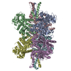
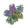
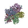
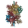
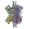
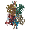
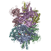
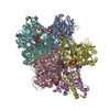
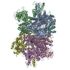
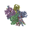
- PDB-6dhl: Bovine glutamate dehydrogenase complexed with epicatechin-3-galla... -
+ Open data

- Basic information
| Entry | Database: PDB / ID: 6dhl | |||||||||
|---|---|---|---|---|---|---|---|---|---|---|
| Title | Bovine glutamate dehydrogenase complexed with epicatechin-3-gallate (ECG) | |||||||||
 Components Components | Glutamate dehydrogenase 1, mitochondrial | |||||||||
 Keywords Keywords | OXIDOREDUCTASE / ECG / glutamate / dehydrogenase / inhibition | |||||||||
| Function / homology |  Function and homology information Function and homology informationGlutamate and glutamine metabolism / Transcriptional activation of mitochondrial biogenesis / L-glutamate dehydrogenase [NAD(P)+] activity / tricarboxylic acid metabolic process / glutamate dehydrogenase [NAD(P)+] / L-glutamate dehydrogenase (NADP+) activity / L-glutamate dehydrogenase (NAD+) activity / L-glutamate catabolic process / L-glutamine metabolic process / Mitochondrial protein degradation ...Glutamate and glutamine metabolism / Transcriptional activation of mitochondrial biogenesis / L-glutamate dehydrogenase [NAD(P)+] activity / tricarboxylic acid metabolic process / glutamate dehydrogenase [NAD(P)+] / L-glutamate dehydrogenase (NADP+) activity / L-glutamate dehydrogenase (NAD+) activity / L-glutamate catabolic process / L-glutamine metabolic process / Mitochondrial protein degradation / positive regulation of insulin secretion / mitochondrial inner membrane / mitochondrial matrix / GTP binding / endoplasmic reticulum / mitochondrion / ATP binding / identical protein binding Similarity search - Function | |||||||||
| Biological species |  | |||||||||
| Method |  X-RAY DIFFRACTION / SYNCHROTRON / MOLECULAR REPLACEMENT / Resolution: 3.624 Å X-RAY DIFFRACTION / SYNCHROTRON / MOLECULAR REPLACEMENT / Resolution: 3.624 Å | |||||||||
 Authors Authors | Smith, T.J. | |||||||||
 Citation Citation | Journal: J. Biol. Chem. / Year: 2011 Title: Green tea polyphenols control dysregulated glutamate dehydrogenase in transgenic mice by hijacking the ADP activation site. Authors: Li, C. / Li, M. / Chen, P. / Narayan, S. / Matschinsky, F.M. / Bennett, M.J. / Stanley, C.A. / Smith, T.J. | |||||||||
| History |
|
- Structure visualization
| Structure viewer | Molecule: MolmilJmol/JSmol |
|---|
- Downloads & links
-Download
| PDBx/mmCIF format | 6dhl.cif.gz | 1.1 MB | Display | PDBx/mmCIF format |
|---|---|---|---|---|
| PDB format | pdb6dhl.ent.gz | 945.2 KB | Display | PDB format |
| PDBx/mmJSON format | 6dhl.json.gz | Tree view | PDBx/mmJSON format | |
| Others |  Other downloads Other downloads |
-Validation report
| Arichive directory | https://data.pdbj.org/pub/pdb/validation_reports/dh/6dhlftp://data.pdbj.org/pub/pdb/validation_reports/dh/6dhl | HTTPS FTP |
|---|
-Related structure data
| Similar structure data |
|---|
-Links
 PDBj
PDBj
- Assembly
| Deposited unit | 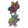
| ||||||||
|---|---|---|---|---|---|---|---|---|---|
| 1 | 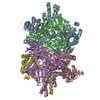
| ||||||||
| 2 | 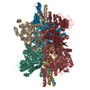
| ||||||||
| Unit cell |
|
-Components
| #1: Protein | Mass: 55127.648 Da / Num. of mol.: 12 / Source method: isolated from a natural source / Source: (natural) References: UniProt: A0A140T871, UniProt: P00366*PLUS, glutamate dehydrogenase (NAD+) #2: Chemical | ChemComp-XEG / ( 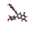  Mass: 442.372 Da / Num. of mol.: 12 / Source method: obtained synthetically / Formula: C22H18O10 Mass: 442.372 Da / Num. of mol.: 12 / Source method: obtained synthetically / Formula: C22H18O10Has protein modification | Y | |
|---|
-Experimental details
-Experiment
| Experiment | Method: X-RAY DIFFRACTION / Number of used crystals: 1 |
|---|
- Sample preparation
| Crystal | Density Matthews: 2.6 Å3/Da / Density % sol: 52.67 % |
|---|---|
| Crystal grow | Temperature: 295 K / Method: vapor diffusion, sitting drop Details: 5 mg/mL GDH in 0.4 mM ECG, 3 mM NADPH, 20 mM glutamate, 2:1 with reservoir solution (0.9 M sodium chloride, 50 mM TEA-HCl, 5 mM reduced glutathione, 8~9% PEG8000, 1 M 1,6-hexanediol), VAPOR ...Details: 5 mg/mL GDH in 0.4 mM ECG, 3 mM NADPH, 20 mM glutamate, 2:1 with reservoir solution (0.9 M sodium chloride, 50 mM TEA-HCl, 5 mM reduced glutathione, 8~9% PEG8000, 1 M 1,6-hexanediol), VAPOR DIFFUSION, HANGING DROP, temperature 295K |
-Data collection
| Diffraction | Mean temperature: 100 K |
|---|---|
| Diffraction source | Source: SYNCHROTRON / Site: APS  / Beamline: 19-ID / Wavelength: 0.97929 Å / Beamline: 19-ID / Wavelength: 0.97929 Å |
| Detector | Type: ADSC QUANTUM 315 / Detector: CCD / Date: Mar 10, 2009 |
| Radiation | Monochromator: double crystal Si(111) / Protocol: SINGLE WAVELENGTH / Monochromatic (M) / Laue (L): M / Scattering type: x-ray |
| Radiation wavelength | Wavelength: 0.97929 Å / Relative weight: 1 |
| Reflection | Resolution: 3.6→50 Å / Num. obs: 72322 / % possible obs: 95.01 % / Redundancy: 3 % / Net I/σ(I): 14.8 |
| Reflection shell | Resolution: 3.6→3.73 Å |
- Processing
| Software |
| ||||||||||||||||||||||||||||||||||||||||||||||||||||||||||||||||||||||||||||||||||||||||||||||||||||||||||||||||||||||||||||||||||||||||||||||||||||||||||||||||||||||||||||||||||||||||||||||||||||
|---|---|---|---|---|---|---|---|---|---|---|---|---|---|---|---|---|---|---|---|---|---|---|---|---|---|---|---|---|---|---|---|---|---|---|---|---|---|---|---|---|---|---|---|---|---|---|---|---|---|---|---|---|---|---|---|---|---|---|---|---|---|---|---|---|---|---|---|---|---|---|---|---|---|---|---|---|---|---|---|---|---|---|---|---|---|---|---|---|---|---|---|---|---|---|---|---|---|---|---|---|---|---|---|---|---|---|---|---|---|---|---|---|---|---|---|---|---|---|---|---|---|---|---|---|---|---|---|---|---|---|---|---|---|---|---|---|---|---|---|---|---|---|---|---|---|---|---|---|---|---|---|---|---|---|---|---|---|---|---|---|---|---|---|---|---|---|---|---|---|---|---|---|---|---|---|---|---|---|---|---|---|---|---|---|---|---|---|---|---|---|---|---|---|---|---|---|---|
| Refinement | Method to determine structure: MOLECULAR REPLACEMENT / Resolution: 3.624→49.706 Å / SU ML: 0.58 / Cross valid method: FREE R-VALUE / σ(F): 1.36 / Phase error: 34.53
| ||||||||||||||||||||||||||||||||||||||||||||||||||||||||||||||||||||||||||||||||||||||||||||||||||||||||||||||||||||||||||||||||||||||||||||||||||||||||||||||||||||||||||||||||||||||||||||||||||||
| Solvent computation | Shrinkage radii: 0.9 Å / VDW probe radii: 1.11 Å | ||||||||||||||||||||||||||||||||||||||||||||||||||||||||||||||||||||||||||||||||||||||||||||||||||||||||||||||||||||||||||||||||||||||||||||||||||||||||||||||||||||||||||||||||||||||||||||||||||||
| Refinement step | Cycle: LAST / Resolution: 3.624→49.706 Å
| ||||||||||||||||||||||||||||||||||||||||||||||||||||||||||||||||||||||||||||||||||||||||||||||||||||||||||||||||||||||||||||||||||||||||||||||||||||||||||||||||||||||||||||||||||||||||||||||||||||
| Refine LS restraints |
| ||||||||||||||||||||||||||||||||||||||||||||||||||||||||||||||||||||||||||||||||||||||||||||||||||||||||||||||||||||||||||||||||||||||||||||||||||||||||||||||||||||||||||||||||||||||||||||||||||||
| LS refinement shell |
|
