 Movie
Movie Controller

[English] 日本語
 Yorodumi
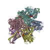
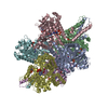
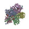
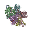
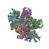
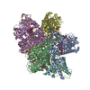
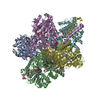
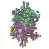
- PDB-1hwy: BOVINE GLUTAMATE DEHYDROGENASE COMPLEXED WITH NAD AND 2-OXOGLUTARATE -
+ Open data

- Basic information
| Entry | Database: PDB / ID: 1hwy | ||||||
|---|---|---|---|---|---|---|---|
| Title | BOVINE GLUTAMATE DEHYDROGENASE COMPLEXED WITH NAD AND 2-OXOGLUTARATE | ||||||
 Components Components | GLUTAMATE DEHYDROGENASE | ||||||
 Keywords Keywords | OXIDOREDUCTASE / allostery / glutamate dehydrogenase / NAD | ||||||
| Function / homology |  Function and homology information Function and homology informationL-glutamate dehydrogenase [NAD(P)+] activity / tricarboxylic acid metabolic process / glutamate dehydrogenase [NAD(P)+] / L-glutamate dehydrogenase (NADP+) activity / L-glutamate dehydrogenase (NAD+) activity / L-glutamate catabolic process / L-glutamine metabolic process / mitochondrial inner membrane / GTP binding / endoplasmic reticulum ...L-glutamate dehydrogenase [NAD(P)+] activity / tricarboxylic acid metabolic process / glutamate dehydrogenase [NAD(P)+] / L-glutamate dehydrogenase (NADP+) activity / L-glutamate dehydrogenase (NAD+) activity / L-glutamate catabolic process / L-glutamine metabolic process / mitochondrial inner membrane / GTP binding / endoplasmic reticulum / mitochondrion / ATP binding / identical protein binding Similarity search - Function | ||||||
| Biological species |  | ||||||
| Method |  X-RAY DIFFRACTION / MOLECULAR REPLACEMENT / Resolution: 3.2 Å X-RAY DIFFRACTION / MOLECULAR REPLACEMENT / Resolution: 3.2 Å | ||||||
 Authors Authors | Smith, T.J. / Peterson, P.E. / Schmidt, T. / Fang, J. / Stanley, C.A. | ||||||
 Citation Citation | Journal: J.Mol.Biol. / Year: 2001 Title: Structures of bovine glutamate dehydrogenase complexes elucidate the mechanism of purine regulation. Authors: Smith, T.J. / Peterson, P.E. / Schmidt, T. / Fang, J. / Stanley, C.A. #1: Journal: Structure / Year: 1999Title: The Structure of Bovine Glutamate Dehydrogenase Provides Insights Into the Mechanism of Allostery Authors: Peterson, P.E. / Smith, T.J. #2: Journal: J.Struct.Biol. / Year: 1997Title: Crystallization and Characterization of Bovine Liver Glutamate Dehydrogenase Authors: Peterson, P.E. / Pierce, J. / Smith, T.J. | ||||||
| History |
|
- Structure visualization
| Structure viewer | Molecule: MolmilJmol/JSmol |
|---|
- Downloads & links
-Download
| PDBx/mmCIF format | 1hwy.cif.gz | 578.9 KB | Display | PDBx/mmCIF format |
|---|---|---|---|---|
| PDB format | pdb1hwy.ent.gz | 483.7 KB | Display | PDB format |
| PDBx/mmJSON format | 1hwy.json.gz | Tree view | PDBx/mmJSON format | |
| Others |  Other downloads Other downloads |
-Validation report
| Arichive directory | https://data.pdbj.org/pub/pdb/validation_reports/hw/1hwyftp://data.pdbj.org/pub/pdb/validation_reports/hw/1hwy | HTTPS FTP |
|---|
-Related structure data
| Related structure data | 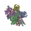 6dhdC 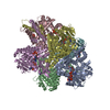 6dhqC  1hwx C: citing same article ( |
|---|---|
| Similar structure data |


-Links
 PDBj
PDBj

- Assembly
| Deposited unit | 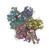
| ||||||||||||||||||||||||||||
|---|---|---|---|---|---|---|---|---|---|---|---|---|---|---|---|---|---|---|---|---|---|---|---|---|---|---|---|---|---|
| 1 |
| ||||||||||||||||||||||||||||
| Unit cell |
| ||||||||||||||||||||||||||||
| Noncrystallographic symmetry (NCS) | NCS oper:
|
-Components
| #1: Protein | Mass: 55638.211 Da / Num. of mol.: 6 / Source method: isolated from a natural source / Source: (natural) References: UniProt: P00366, glutamate dehydrogenase [NAD(P)+] #2: Chemical | ChemComp-PO4 /   Mass: 94.971 Da / Num. of mol.: 24 / Source method: obtained synthetically / Formula: PO4 Mass: 94.971 Da / Num. of mol.: 24 / Source method: obtained synthetically / Formula: PO4#3: Chemical | ChemComp-AKG /   Mass: 146.098 Da / Num. of mol.: 6 / Source method: obtained synthetically / Formula: C5H6O5 Mass: 146.098 Da / Num. of mol.: 6 / Source method: obtained synthetically / Formula: C5H6O5#4: Chemical | ChemComp-NAD /   Mass: 663.425 Da / Num. of mol.: 12 / Source method: obtained synthetically / Formula: C21H27N7O14P2 / Comment: NAD*YM Mass: 663.425 Da / Num. of mol.: 12 / Source method: obtained synthetically / Formula: C21H27N7O14P2 / Comment: NAD*YM#5: Water | ChemComp-HOH / |  Mass: 18.015 Da / Num. of mol.: 36 / Source method: isolated from a natural source / Formula: H2O Mass: 18.015 Da / Num. of mol.: 36 / Source method: isolated from a natural source / Formula: H2O |
|---|
-Experimental details
-Experiment
| Experiment | Method: X-RAY DIFFRACTION / Number of used crystals: 1 |
|---|
- Sample preparation
| Crystal | Density Matthews: 2.98 Å3/Da / Density % sol: 58.73 % | |||||||||||||||||||||||||||||||||||||||||||||||||||||||
|---|---|---|---|---|---|---|---|---|---|---|---|---|---|---|---|---|---|---|---|---|---|---|---|---|---|---|---|---|---|---|---|---|---|---|---|---|---|---|---|---|---|---|---|---|---|---|---|---|---|---|---|---|---|---|---|---|
| Crystal grow | Temperature: 298 K / Method: vapor diffusion, sitting drop / pH: 7 Details: sodium phosphate, NaCl, PEG 8000, sodium azide, methyl pentanediol, octyl-b-glucopyranoside, pH 7, VAPOR DIFFUSION, SITTING DROP, temperature 298K | |||||||||||||||||||||||||||||||||||||||||||||||||||||||
| Crystal grow | *PLUS | |||||||||||||||||||||||||||||||||||||||||||||||||||||||
| Components of the solutions | *PLUS
|
-Data collection
| Diffraction | Mean temperature: 100 K |
|---|---|
| Diffraction source | Source: ROTATING ANODE / Type: RIGAKU RU200 / Wavelength: 1.5418 |
| Detector | Type: RIGAKU RAXIS IV / Detector: IMAGE PLATE / Date: 1999 |
| Radiation | Protocol: SINGLE WAVELENGTH / Monochromatic (M) / Laue (L): M / Scattering type: x-ray |
| Radiation wavelength | Wavelength: 1.5418 Å / Relative weight: 1 |
| Reflection | Resolution: 3→20 Å |
| Reflection shell | Resolution: 3.2→3.4 Å / Rmerge(I) obs: 0.12 / % possible all: 78 |
| Reflection | *PLUS % possible obs: 79 % / Rmerge(I) obs: 0.123 |
| Reflection shell | *PLUS % possible obs: 80 % / Rmerge(I) obs: 0.475 |
- Processing
| Software |
| ||||||||||||||||||||||||||||||
|---|---|---|---|---|---|---|---|---|---|---|---|---|---|---|---|---|---|---|---|---|---|---|---|---|---|---|---|---|---|---|---|
| Refinement | Method to determine structure: MOLECULAR REPLACEMENT / Resolution: 3.2→8 Å / σ(F): 3 /
| ||||||||||||||||||||||||||||||
| Refinement step | Cycle: LAST / Resolution: 3.2→8 Å
| ||||||||||||||||||||||||||||||
| Software | *PLUS Name: X-PLOR / Classification: refinement | ||||||||||||||||||||||||||||||
| Refine LS restraints | *PLUS
|
