 Movie
Movie Controller

[English] 日本語
 Yorodumi
- PDB-5upn: Crystal structure of BhGH81 mutant in complex with laminaro-tetraose -
+ Open data

- Basic information
| Entry | Database: PDB / ID: 5upn | |||||||||
|---|---|---|---|---|---|---|---|---|---|---|
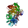
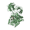
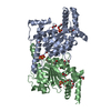
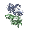
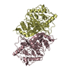
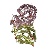
| Title | Crystal structure of BhGH81 mutant in complex with laminaro-tetraose | |||||||||
 Components Components | BH0236 protein | |||||||||
 Keywords Keywords | HYDROLASE / (alpha/beta)6 barrel / glycoside hydrolase | |||||||||
| Function / homology |  Function and homology information Function and homology informationendo-1,3(4)-beta-glucanase activity / glucan endo-1,3-beta-D-glucosidase / glucan endo-1,3-beta-D-glucosidase activity / polysaccharide catabolic process / cell wall organization / carbohydrate binding / extracellular region Similarity search - Function | |||||||||
| Biological species |  Bacillus halodurans (bacteria) Bacillus halodurans (bacteria) | |||||||||
| Method |  X-RAY DIFFRACTION / SYNCHROTRON / MOLECULAR REPLACEMENT / molecular replacement / Resolution: 1.8 Å X-RAY DIFFRACTION / SYNCHROTRON / MOLECULAR REPLACEMENT / molecular replacement / Resolution: 1.8 Å | |||||||||
 Authors Authors | Pluvinage, B. / Boraston, A.B. | |||||||||
 Citation Citation | Journal: Structure / Year: 2017 Title: The quaternary structure of beta-1,3-glucan contributes to its recognition and hydrolysis by a multimodular family 81 glycoside hydrolase Authors: Pluvinage, B. / Fillo, A. / Massel, P. / Boraston, A.B. | |||||||||
| History |
|
- Structure visualization
| Structure viewer | Molecule: MolmilJmol/JSmol |
|---|
- Downloads & links
-Download
| PDBx/mmCIF format | 5upn.cif.gz | 190.8 KB | Display | PDBx/mmCIF format |
|---|---|---|---|---|
| PDB format | pdb5upn.ent.gz | 146.2 KB | Display | PDB format |
| PDBx/mmJSON format | 5upn.json.gz | Tree view | PDBx/mmJSON format | |
| Others |  Other downloads Other downloads |
-Validation report
| Arichive directory | https://data.pdbj.org/pub/pdb/validation_reports/up/5upnftp://data.pdbj.org/pub/pdb/validation_reports/up/5upn | HTTPS FTP |
|---|
-Related structure data
| Related structure data | 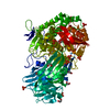 5upiC  5upmC 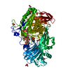 5upoC 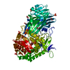 5v1wC  5t49S C: citing same article ( S: Starting model for refinement |
|---|---|
| Similar structure data |


-Links
 PDBj
PDBj


- Assembly
| Deposited unit | 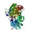
| ||||||||
|---|---|---|---|---|---|---|---|---|---|
| 1 |
| ||||||||
| Unit cell |
|
-Components
-Protein , 1 types, 1 molecules A
| #1: Protein | Mass: 85277.633 Da / Num. of mol.: 1 / Fragment: residues 28-779 / Mutation: E542Q Source method: isolated from a genetically manipulated source Source: (gene. exp.) Bacillus halodurans (strain ATCC BAA-125 / DSM 18197 / FERM 7344 / JCM 9153 / C-125) (bacteria)Strain: ATCC BAA-125 / DSM 18197 / FERM 7344 / JCM 9153 / C-125 Gene: BH0236 / Plasmid: pET28a / Production host: |
|---|
-Sugars , 4 types, 6 molecules 

| #2: Polysaccharide | 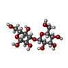  Source method: isolated from a genetically manipulated source Details: oligosaccharide / References: beta-laminaribiose #3: Polysaccharide | beta-D-glucopyranose-(1-3)-beta-D-glucopyranose-(1-3)-beta-D-glucopyranose-(1-3)-beta-D-glucopyranose | Source method: isolated from a genetically manipulated source #4: Sugar | ChemComp-GLC / |  Type: D-saccharide, alpha linking / Mass: 180.156 Da / Num. of mol.: 1 Type: D-saccharide, alpha linking / Mass: 180.156 Da / Num. of mol.: 1Source method: isolated from a genetically manipulated source Formula: C6H12O6 #5: Sugar |  Type: D-saccharide, beta linking / Mass: 180.156 Da / Num. of mol.: 2 Type: D-saccharide, beta linking / Mass: 180.156 Da / Num. of mol.: 2Source method: isolated from a genetically manipulated source Formula: C6H12O6 |
|---|
-Non-polymers , 3 types, 824 molecules 


| #6: Chemical | ChemComp-PO4 /  Mass: 94.971 Da / Num. of mol.: 9 / Source method: obtained synthetically / Formula: PO4 Mass: 94.971 Da / Num. of mol.: 9 / Source method: obtained synthetically / Formula: PO4#7: Chemical | ChemComp-EDO /  Mass: 62.068 Da / Num. of mol.: 7 / Source method: obtained synthetically / Formula: C2H6O2 Mass: 62.068 Da / Num. of mol.: 7 / Source method: obtained synthetically / Formula: C2H6O2#8: Water | ChemComp-HOH / | Mass: 18.015 Da / Num. of mol.: 808 / Source method: isolated from a natural source / Formula: H2O |
|---|
-Experimental details
-Experiment
| Experiment | Method: X-RAY DIFFRACTION / Number of used crystals: 1 |
|---|
- Sample preparation
| Crystal | Density Matthews: 2.91 Å3/Da / Density % sol: 57.71 % |
|---|---|
| Crystal grow | Temperature: 291 K / Method: vapor diffusion, hanging drop / pH: 5.5 Details: 15% PEG 10000, 0.2 M ammonium acetate, 0.1 M bis-tris |
-Data collection
| Diffraction | Mean temperature: 100 K |
|---|---|
| Diffraction source | Source: SYNCHROTRON / Site: CLSI  / Beamline: 08ID-1 / Wavelength: 0.97971 Å / Beamline: 08ID-1 / Wavelength: 0.97971 Å |
| Detector | Type: RAYONIX MX-300 / Detector: CCD / Date: Sep 9, 2014 |
| Radiation | Protocol: SINGLE WAVELENGTH / Monochromatic (M) / Laue (L): M / Scattering type: x-ray |
| Radiation wavelength | Wavelength: 0.97971 Å / Relative weight: 1 |
| Reflection | Resolution: 1.8→79.14 Å / Num. obs: 89565 / % possible obs: 96.7 % / Redundancy: 6.7 % / CC1/2: 0.997 / Rmerge(I) obs: 0.09 / Rpim(I) all: 0.036 / Net I/σ(I): 13.7 |
| Reflection shell | Resolution: 1.8→1.9 Å / Redundancy: 6 % / Rmerge(I) obs: 0.276 / Mean I/σ(I) obs: 5.6 / CC1/2: 0.949 / Rpim(I) all: 0.116 / % possible all: 91.8 |
-Phasing
| Phasing | Method: molecular replacement |
|---|
- Processing
| Software |
| ||||||||||||||||||||||||||||||||||||||||||||||||||||||||||||
|---|---|---|---|---|---|---|---|---|---|---|---|---|---|---|---|---|---|---|---|---|---|---|---|---|---|---|---|---|---|---|---|---|---|---|---|---|---|---|---|---|---|---|---|---|---|---|---|---|---|---|---|---|---|---|---|---|---|---|---|---|---|
| Refinement | Method to determine structure: MOLECULAR REPLACEMENT Starting model: 5T49 Resolution: 1.8→79.14 Å / Cor.coef. Fo:Fc: 0.966 / Cor.coef. Fo:Fc free: 0.961 / SU B: 1.911 / SU ML: 0.059 / Cross valid method: THROUGHOUT / σ(F): 0 / ESU R: 0.098 / ESU R Free: 0.09 Details: HYDROGENS HAVE BEEN ADDED IN THE RIDING POSITIONS U VALUES : REFINED INDIVIDUALLY
| ||||||||||||||||||||||||||||||||||||||||||||||||||||||||||||
| Solvent computation | Ion probe radii: 0.8 Å / Shrinkage radii: 0.8 Å / VDW probe radii: 1.2 Å | ||||||||||||||||||||||||||||||||||||||||||||||||||||||||||||
| Displacement parameters | Biso max: 60.96 Å2 / Biso mean: 16.5 Å2 / Biso min: 2.66 Å2
| ||||||||||||||||||||||||||||||||||||||||||||||||||||||||||||
| Refinement step | Cycle: final / Resolution: 1.8→79.14 Å
| ||||||||||||||||||||||||||||||||||||||||||||||||||||||||||||
| Refine LS restraints |
| ||||||||||||||||||||||||||||||||||||||||||||||||||||||||||||
| LS refinement shell | Resolution: 1.8→1.847 Å / Rfactor Rfree error: 0 / Total num. of bins used: 20
|
