 Movie
Movie Controller

[English] 日本語
 Yorodumi
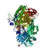
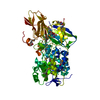
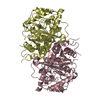
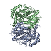
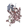
- PDB-5xlw: Mycobacterium tuberculosis Pantothenate kinase mutant F247A/F254A -
+ Open data

- Basic information
| Entry | Database: PDB / ID: 5xlw | ||||||
|---|---|---|---|---|---|---|---|
| Title | Mycobacterium tuberculosis Pantothenate kinase mutant F247A/F254A | ||||||
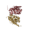
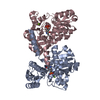
 Components Components | Pantothenate kinase | ||||||
 Keywords Keywords | TRANSFERASE / Homodimer / CoA biosynthesis / Nucleotide binding / Concerted movement / Structural transformation | ||||||
| Function / homology |  Function and homology information Function and homology informationpantothenate kinase / pantothenate kinase activity / coenzyme A biosynthetic process / ATP binding / plasma membrane / cytoplasm Similarity search - Function | ||||||
| Biological species |   Mycobacterium tuberculosis (bacteria) Mycobacterium tuberculosis (bacteria) | ||||||
| Method |  X-RAY DIFFRACTION / SYNCHROTRON / MOLECULAR REPLACEMENT / Resolution: 2.26 Å X-RAY DIFFRACTION / SYNCHROTRON / MOLECULAR REPLACEMENT / Resolution: 2.26 Å | ||||||
 Authors Authors | Paul, A. / Kumar, P. / Surolia, A. / Vijayan, M. | ||||||
 Citation Citation | Journal: Acta Crystallogr F Struct Biol Commun / Year: 2017 Title: Biochemical and structural studies of mutants indicate concerted movement of the dimer interface and ligand-binding region of Mycobacterium tuberculosis pantothenate kinase Authors: Paul, A. / Kumar, P. / Surolia, A. / Vijayan, M. #1: Journal: Acta Crystallogr. Sect. F Struct. Biol. Cryst. Commun. Year: 2005 Title: Expression, purification, crystallization and preliminary X-ray crystallographic analysis of pantothenate kinase from Mycobacterium tuberculosis. Authors: Das, S. / Kumar, P. / Bhor, V. / Surolia, A. / Vijayan, M. #2: Journal: Acta Crystallogr. D Biol. Crystallogr. / Year: 2006Title: Invariance and variability in bacterial PanK: a study based on the crystal structure of Mycobacterium tuberculosis PanK. Authors: Das, S. / Kumar, P. / Bhor, V. / Surolia, A. / Vijayan, M. #3: Journal: Acta Crystallogr. D Biol. Crystallogr. / Year: 2009Title: Mycobacterium tuberculosis pantothenate kinase: possible changes in location of ligands during enzyme action. Authors: Chetnani, B. / Das, S. / Kumar, P. / Surolia, A. / Vijayan, M. #4: Journal: J. Mol. Biol. / Year: 2010Title: M. tuberculosis pantothenate kinase: dual substrate specificity and unusual changes in ligand locations. Authors: Chetnani, B. / Kumar, P. / Surolia, A. / Vijayan, M. #5: Journal: Acta Crystallogr. D Biol. Crystallogr. / Year: 2011Title: Location and conformation of pantothenate and its derivatives in Mycobacterium tuberculosis pantothenate kinase: insights into enzyme action. Authors: Chetnani, B. / Kumar, P. / Abhinav, K.V. / Chhibber, M. / Surolia, A. / Vijayan, M. | ||||||
| History |
|
- Structure visualization
| Structure viewer | Molecule: MolmilJmol/JSmol |
|---|
- Downloads & links
-Download
| PDBx/mmCIF format | 5xlw.cif.gz | 136.8 KB | Display | PDBx/mmCIF format |
|---|---|---|---|---|
| PDB format | pdb5xlw.ent.gz | 106.5 KB | Display | PDB format |
| PDBx/mmJSON format | 5xlw.json.gz | Tree view | PDBx/mmJSON format | |
| Others |  Other downloads Other downloads |
-Validation report
| Arichive directory | https://data.pdbj.org/pub/pdb/validation_reports/xl/5xlwftp://data.pdbj.org/pub/pdb/validation_reports/xl/5xlw | HTTPS FTP |
|---|
-Related structure data
| Related structure data |  5xlvC  5xmbC 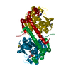 2geuS C: citing same article ( S: Starting model for refinement |
|---|---|
| Similar structure data |


-Links
 PDBj
PDBj- Assembly
| Deposited unit | 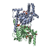
| ||||||||
|---|---|---|---|---|---|---|---|---|---|
| 1 |
| ||||||||
| Unit cell |
|
-Components
-Protein , 1 types, 2 molecules BA
| #1: Protein | Mass: 35552.660 Da / Num. of mol.: 2 / Mutation: F247A, F254A Source method: isolated from a genetically manipulated source Source: (gene. exp.) Mycobacterium tuberculosis (strain ATCC 25618 / H37Rv) (bacteria)Strain: ATCC 25618 / H37Rv / Gene: coaA, Rv1092c, MTV017.45c / Plasmid: Pet28a(+) / Details (production host): Novagen / Production host: |
|---|
-Non-polymers , 5 types, 204 molecules 


| #2: Chemical | ChemComp-EDO /  Mass: 62.068 Da / Num. of mol.: 20 / Source method: obtained synthetically / Formula: C2H6O2 Mass: 62.068 Da / Num. of mol.: 20 / Source method: obtained synthetically / Formula: C2H6O2#3: Chemical | ChemComp-1PE / |  Mass: 238.278 Da / Num. of mol.: 1 / Source method: obtained synthetically / Formula: C10H22O6 / Comment: precipitant*YM Mass: 238.278 Da / Num. of mol.: 1 / Source method: obtained synthetically / Formula: C10H22O6 / Comment: precipitant*YM#4: Chemical | ChemComp-SO4 /  Mass: 96.063 Da / Num. of mol.: 4 / Source method: obtained synthetically / Formula: SO4 Mass: 96.063 Da / Num. of mol.: 4 / Source method: obtained synthetically / Formula: SO4#5: Chemical | ChemComp-PEG / |  Mass: 106.120 Da / Num. of mol.: 1 / Source method: obtained synthetically / Formula: C4H10O3 Mass: 106.120 Da / Num. of mol.: 1 / Source method: obtained synthetically / Formula: C4H10O3#6: Water | ChemComp-HOH / | Mass: 18.015 Da / Num. of mol.: 178 / Source method: isolated from a natural source / Formula: H2O |
|---|
-Experimental details
-Experiment
| Experiment | Method: X-RAY DIFFRACTION / Number of used crystals: 1 |
|---|
- Sample preparation
| Crystal | Density Matthews: 3.68 Å3/Da / Density % sol: 66.61 % Description: THE ENTRY CONTAINS FRIEDEL PAIRS IN F_PLUS/MINUS COLUMNS. |
|---|---|
| Crystal grow | Temperature: 295 K / Method: microbatch / pH: 7.5 Details: 2% PEG 400, 2.0 M Ammonium sulphate, 100mM HEPES sodium salt |
-Data collection
| Diffraction | Mean temperature: 100 K |
|---|---|
| Diffraction source | Source: SYNCHROTRON / Site: ESRF  / Beamline: BM14 / Wavelength: 0.95372 Å / Beamline: BM14 / Wavelength: 0.95372 Å |
| Detector | Type: MARMOSAIC 225 mm CCD / Detector: CCD / Date: Jul 31, 2012 |
| Radiation | Protocol: SINGLE WAVELENGTH / Monochromatic (M) / Laue (L): M / Scattering type: x-ray |
| Radiation wavelength | Wavelength: 0.95372 Å / Relative weight: 1 |
| Reflection | Resolution: 2.26→43.55 Å / Num. obs: 49403 / % possible obs: 99.7 % / Redundancy: 7.5 % / Rmerge(I) obs: 0.082 / Net I/σ(I): 15.5 |
| Reflection shell | Resolution: 2.26→2.38 Å / Redundancy: 7.2 % / Rmerge(I) obs: 0.498 / Mean I/σ(I) obs: 3.7 / Num. unique obs: 7188 / % possible all: 100 |
- Processing
| Software |
| ||||||||||||||||||||||||||||||||||||||||||||||||||||||||||||||||||||||||||||||||||||||||||||||||||||||||||||||||||||||||||||||||||||||||||||||||||||||||||||||||||||||||||||||||||||||
|---|---|---|---|---|---|---|---|---|---|---|---|---|---|---|---|---|---|---|---|---|---|---|---|---|---|---|---|---|---|---|---|---|---|---|---|---|---|---|---|---|---|---|---|---|---|---|---|---|---|---|---|---|---|---|---|---|---|---|---|---|---|---|---|---|---|---|---|---|---|---|---|---|---|---|---|---|---|---|---|---|---|---|---|---|---|---|---|---|---|---|---|---|---|---|---|---|---|---|---|---|---|---|---|---|---|---|---|---|---|---|---|---|---|---|---|---|---|---|---|---|---|---|---|---|---|---|---|---|---|---|---|---|---|---|---|---|---|---|---|---|---|---|---|---|---|---|---|---|---|---|---|---|---|---|---|---|---|---|---|---|---|---|---|---|---|---|---|---|---|---|---|---|---|---|---|---|---|---|---|---|---|---|---|
| Refinement | Method to determine structure: MOLECULAR REPLACEMENT Starting model: 2GEU Resolution: 2.26→43.55 Å / Cor.coef. Fo:Fc: 0.952 / Cor.coef. Fo:Fc free: 0.933 / SU B: 5.504 / SU ML: 0.133 / Cross valid method: THROUGHOUT / ESU R: 0.188 / ESU R Free: 0.171 / Stereochemistry target values: MAXIMUM LIKELIHOOD / Details: HYDROGENS HAVE BEEN ADDED IN THE RIDING POSITIONS
| ||||||||||||||||||||||||||||||||||||||||||||||||||||||||||||||||||||||||||||||||||||||||||||||||||||||||||||||||||||||||||||||||||||||||||||||||||||||||||||||||||||||||||||||||||||||
| Solvent computation | Ion probe radii: 0.8 Å / Shrinkage radii: 0.8 Å / VDW probe radii: 1.2 Å / Solvent model: MASK | ||||||||||||||||||||||||||||||||||||||||||||||||||||||||||||||||||||||||||||||||||||||||||||||||||||||||||||||||||||||||||||||||||||||||||||||||||||||||||||||||||||||||||||||||||||||
| Displacement parameters | Biso mean: 41.687 Å2
| ||||||||||||||||||||||||||||||||||||||||||||||||||||||||||||||||||||||||||||||||||||||||||||||||||||||||||||||||||||||||||||||||||||||||||||||||||||||||||||||||||||||||||||||||||||||
| Refinement step | Cycle: 1 / Resolution: 2.26→43.55 Å
| ||||||||||||||||||||||||||||||||||||||||||||||||||||||||||||||||||||||||||||||||||||||||||||||||||||||||||||||||||||||||||||||||||||||||||||||||||||||||||||||||||||||||||||||||||||||
| Refine LS restraints |
|