- PDB-3d3a: Crystal structure of a beta-galactosidase from Bacteroides thetai... -
+
Open data
ID or keywords:
Loading...
-
Basic information
Entry
Database: PDB / ID: 3d3a
Title
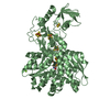
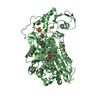
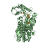
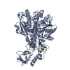
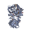
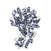
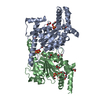
Crystal structure of a beta-galactosidase from Bacteroides thetaiotaomicron
Components
Beta-galactosidase
Keywords
HYDROLASE / beta-galactosidase / Bacteroides thetaiotaomicron / Protein Structure Initiative II / PSI II / NYSGXRC / 11092f / Structural Genomics / New York SGX Research Center for Structural Genomics / Glycosidase
Function / homology
Function and homology information
galactose catabolic process / beta-galactosidase / beta-galactosidase activity / vacuole / metal ion binding Similarity search - Function
: / Beta-galactosidase, first all-beta domain / : / Beta-galactosidase, galactose-binding domain / Glycoside hydrolase 35, catalytic domain / Glycosyl hydrolases family 35 / Glycoside hydrolase, family 35 / Coagulation factors 5/8 type C domain (FA58C) profile. / F5/8 type C domain / Coagulation factor 5/8 C-terminal domain ...: / Beta-galactosidase, first all-beta domain / : / Beta-galactosidase, galactose-binding domain / Glycoside hydrolase 35, catalytic domain / Glycosyl hydrolases family 35 / Glycoside hydrolase, family 35 / Coagulation factors 5/8 type C domain (FA58C) profile. / F5/8 type C domain / Coagulation factor 5/8 C-terminal domain / Galactose-binding domain-like / Galactose-binding-like domain superfamily / Glycosidases / Prokaryotic membrane lipoprotein lipid attachment site profile. / Glycoside hydrolase superfamily / Jelly Rolls / TIM Barrel / Alpha-Beta Barrel / Sandwich / Mainly Beta / Alpha Beta Similarity search - Domain/homology
Resolution: 2.13→2.21 Å / Redundancy: 2 % / Rmerge(I) obs: 0.293 / Mean I/σ(I) obs: 1 / Num. unique all: 2607 / % possible all: 57.6
-
Processing
Software
Name
Version
Classification
CNS
1.1
refinement
CBASS
datacollection
HKL-2000
datareduction
HKL-2000
datascaling
SHELXD
phasing
SHARP
phasing
ARP/wARP
modelbuilding
Refinement
Method to determine structure: SAD / Resolution: 2.15→32.46 Å / Rfactor Rfree error: 0.006 / Data cutoff high absF: 51761.57 / Data cutoff low absF: 0 / Isotropic thermal model: RESTRAINED / Cross valid method: THROUGHOUT / σ(F): 0 / Stereochemistry target values: Engh & Huber Details: Residues listed as missing in Remark 465 are due to lack of electron density. Residues with missing atoms listed in Remark 470 are due to lack of electron density for side chains and modeled as alanines.
In the structure databanks used in Yorodumi, some data are registered as the other names, "COVID-19 virus" and "2019-nCoV". Here are the details of the virus and the list of structure data.
Jan 31, 2019. EMDB accession codes are about to change! (news from PDBe EMDB page)
EMDB accession codes are about to change! (news from PDBe EMDB page)
The allocation of 4 digits for EMDB accession codes will soon come to an end. Whilst these codes will remain in use, new EMDB accession codes will include an additional digit and will expand incrementally as the available range of codes is exhausted. The current 4-digit format prefixed with “EMD-” (i.e. EMD-XXXX) will advance to a 5-digit format (i.e. EMD-XXXXX), and so on. It is currently estimated that the 4-digit codes will be depleted around Spring 2019, at which point the 5-digit format will come into force.
The EM Navigator/Yorodumi systems omit the EMD- prefix.
Related info.:Q: What is EMD? / ID/Accession-code notation in Yorodumi/EM Navigator
Yorodumi is a browser for structure data from EMDB, PDB, SASBDB, etc.
This page is also the successor to EM Navigator detail page, and also detail information page/front-end page for Omokage search.
The word "yorodu" (or yorozu) is an old Japanese word meaning "ten thousand". "mi" (miru) is to see.
Related info.:EMDB / PDB / SASBDB / Comparison of 3 databanks / Yorodumi Search / Aug 31, 2016. New EM Navigator & Yorodumi / Yorodumi Papers / Jmol/JSmol / Function and homology information / Changes in new EM Navigator and Yorodumi
 Movie
Movie Controller

 Yorodumi
 Open data
 Basic information
 Components
 Keywords
 Function and homology information
 Bacteroides thetaiotaomicron VPI-5482 (bacteria)
 X-RAY DIFFRACTION /
  Authors
 Citation
 Structure visualization
 Downloads & links
 Other downloads
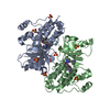


 PDBj
PDBj


 Assembly
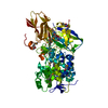
 Mass: 18.015 Da / Num. of mol.: 384 / Source method: isolated from a natural source / Formula: H2O
 Sample preparation
 / Beamline: X29A / Wavelength: 0.979 Å
 Processing