 Movie
Movie Controller

[English] 日本語
 Yorodumi
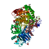
- PDB-1l8x: Crystal Structure of Ferrochelatase from the Yeast, Saccharomyces... -
+ Open data

- Basic information
| Entry | Database: PDB / ID: 1l8x | ||||||
|---|---|---|---|---|---|---|---|
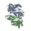
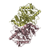
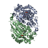
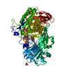
| Title | Crystal Structure of Ferrochelatase from the Yeast, Saccharomyces cerevisiae, with Cobalt(II) as the Substrate Ion | ||||||
 Components Components | Ferrochelatase | ||||||
 Keywords Keywords | LYASE / Ferrochelatase / Heme Biosynthesis / protoheme / Ferro-Lyase / Porphyrin Metallation / Cobalt / Mitochondrial inner membrane protein | ||||||
| Function / homology |  Function and homology information Function and homology informationHeme biosynthesis / protoporphyrin ferrochelatase / protoporphyrin ferrochelatase activity / Mitochondrial protein degradation / heme biosynthetic process / mitochondrial inner membrane / mitochondrion Similarity search - Function | ||||||
| Biological species |  | ||||||
| Method |  X-RAY DIFFRACTION / SYNCHROTRON / MOLECULAR REPLACEMENT / Resolution: 2.7 Å X-RAY DIFFRACTION / SYNCHROTRON / MOLECULAR REPLACEMENT / Resolution: 2.7 Å | ||||||
 Authors Authors | Karlberg, T. / Lecerof, D. / Gora, M. / Silvegren, G. / Labbe-Bois, R. / Hansson, M. / Al-Karadaghi, S. | ||||||
 Citation Citation | Journal: Biochemistry / Year: 2002 Title: Metal Binding to Saccharomyces cerevisiae Ferrochelatase Authors: Karlberg, T. / Lecerof, D. / Gora, M. / Silvegren, G. / Labbe-Bois, R. / Hansson, M. / Al-Karadaghi, S. | ||||||
| History |
|
- Structure visualization
| Structure viewer | Molecule: MolmilJmol/JSmol |
|---|
- Downloads & links
-Download
| PDBx/mmCIF format | 1l8x.cif.gz | 149.7 KB | Display | PDBx/mmCIF format |
|---|---|---|---|---|
| PDB format | pdb1l8x.ent.gz | 117.5 KB | Display | PDB format |
| PDBx/mmJSON format | 1l8x.json.gz | Tree view | PDBx/mmJSON format | |
| Others |  Other downloads Other downloads |
-Validation report
| Arichive directory | https://data.pdbj.org/pub/pdb/validation_reports/l8/1l8xftp://data.pdbj.org/pub/pdb/validation_reports/l8/1l8x | HTTPS FTP |
|---|
-Related structure data
| Related structure data |  1lbqC  1hrkS S: Starting model for refinement C: citing same article ( |
|---|---|
| Similar structure data |


-Links
 PDBj
PDBj

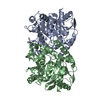
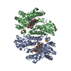
- Assembly
| Deposited unit | 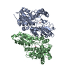
| ||||||||
|---|---|---|---|---|---|---|---|---|---|
| 1 |
| ||||||||
| Unit cell |
|
-Components
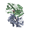
| #1: Protein | Mass: 41000.727 Da / Num. of mol.: 2 Source method: isolated from a genetically manipulated source Source: (gene. exp.) Gene: HEMZ / Production host:  #2: Chemical |   Mass: 58.933 Da / Num. of mol.: 2 / Source method: obtained synthetically / Formula: Co Mass: 58.933 Da / Num. of mol.: 2 / Source method: obtained synthetically / Formula: Co#3: Water | ChemComp-HOH / |  Mass: 18.015 Da / Num. of mol.: 9 / Source method: isolated from a natural source / Formula: H2O Mass: 18.015 Da / Num. of mol.: 9 / Source method: isolated from a natural source / Formula: H2O |
|---|
-Experimental details
-Experiment
| Experiment | Method: X-RAY DIFFRACTION / Number of used crystals: 1 |
|---|
- Sample preparation
| Crystal | Density Matthews: 3 Å3/Da / Density % sol: 59 % | ||||||||||||||||||||||||||||||||||||||||||
|---|---|---|---|---|---|---|---|---|---|---|---|---|---|---|---|---|---|---|---|---|---|---|---|---|---|---|---|---|---|---|---|---|---|---|---|---|---|---|---|---|---|---|---|
| Crystal grow | Temperature: 277 K / Method: vapor diffusion, hanging drop / pH: 7.5 Details: PEG2000, 2-propanol, Tris-HCl, pH 7.5, VAPOR DIFFUSION, HANGING DROP, temperature 277K | ||||||||||||||||||||||||||||||||||||||||||
| Crystal grow | *PLUS Temperature: 4 ℃ / pH: 8 | ||||||||||||||||||||||||||||||||||||||||||
| Components of the solutions | *PLUS
|
-Data collection
| Diffraction | Mean temperature: 100 K |
|---|---|
| Diffraction source | Source: SYNCHROTRON / Site: MAX II  / Beamline: I711 / Wavelength: 0.9678 Å / Beamline: I711 / Wavelength: 0.9678 Å |
| Detector | Type: MARRESEARCH / Detector: CCD / Date: Nov 11, 2001 |
| Radiation | Monochromator: Mirrors / Protocol: SINGLE WAVELENGTH / Monochromatic (M) / Laue (L): M / Scattering type: x-ray |
| Radiation wavelength | Wavelength: 0.9678 Å / Relative weight: 1 |
| Reflection | Resolution: 2.7→15 Å / Num. all: 22973 / Num. obs: 22973 / % possible obs: 90.9 % / Observed criterion σ(F): 0 / Observed criterion σ(I): 0 |
| Reflection shell | Resolution: 2.7→2.9 Å / Rmerge(I) obs: 0.302 / Mean I/σ(I) obs: 22.9 / Num. unique all: 1710 / % possible all: 60.9 |
| Reflection | *PLUS Lowest resolution: 15 Å / Rmerge(I) obs: 0.174 |
| Reflection shell | *PLUS % possible obs: 60.9 % / Mean I/σ(I) obs: 0.302 |
- Processing
| Software |
| |||||||||||||||||||||||||||||||||||
|---|---|---|---|---|---|---|---|---|---|---|---|---|---|---|---|---|---|---|---|---|---|---|---|---|---|---|---|---|---|---|---|---|---|---|---|---|
| Refinement | Method to determine structure: MOLECULAR REPLACEMENT Starting model: PDB ENTRY 1HRK Resolution: 2.7→15 Å / Cross valid method: THROUGHOUT USING FREE R / σ(F): 0 / σ(I): 0 / Stereochemistry target values: Engh & Huber
| |||||||||||||||||||||||||||||||||||
| Refine analyze |
| |||||||||||||||||||||||||||||||||||
| Refinement step | Cycle: LAST / Resolution: 2.7→15 Å
| |||||||||||||||||||||||||||||||||||
| Refine LS restraints |
| |||||||||||||||||||||||||||||||||||
| LS refinement shell |
| |||||||||||||||||||||||||||||||||||
| Refinement | *PLUS Lowest resolution: 15 Å / % reflection Rfree: 5 % | |||||||||||||||||||||||||||||||||||
| Solvent computation | *PLUS | |||||||||||||||||||||||||||||||||||
| Displacement parameters | *PLUS | |||||||||||||||||||||||||||||||||||
| Refine LS restraints | *PLUS
|
