 Movie
Movie Controller

[English] 日本語
 Yorodumi
- PDB-1obj: Crystal structure of the T150A mutant of Malonamidase E2 from Bra... -
+ Open data

- Basic information
| Entry | Database: PDB / ID: 1obj | ||||||
|---|---|---|---|---|---|---|---|
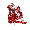
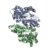
| Title | Crystal structure of the T150A mutant of Malonamidase E2 from Bradyrhizobium japonicum | ||||||
 Components Components | MALONAMIDASE E2 | ||||||
 Keywords Keywords | AMIDASE | ||||||
| Function / homology |  Function and homology information Function and homology information | ||||||
| Biological species |  BRADYRHIZOBIUM JAPONICUM (bacteria) BRADYRHIZOBIUM JAPONICUM (bacteria) | ||||||
| Method |  X-RAY DIFFRACTION / DIRECT METHODS / Resolution: 1.9 Å X-RAY DIFFRACTION / DIRECT METHODS / Resolution: 1.9 Å | ||||||
 Authors Authors | Shin, S. / Oh, B.-H. | ||||||
 Citation Citation | Journal: J.Biol.Chem. / Year: 2003 Title: Characterization of a Novel Ser-Cisser-Lys Catalytic Triad in Comparison with the Classical Ser-His-Asp Triad Authors: Shin, S. / Yun, Y.S. / Koo, H.M. / Kim, Y.S. / Choi, K.Y. / Oh, B.-H. | ||||||
| History |
|
- Structure visualization
| Structure viewer | Molecule: MolmilJmol/JSmol |
|---|
- Downloads & links
-Download
| PDBx/mmCIF format | 1obj.cif.gz | 179.1 KB | Display | PDBx/mmCIF format |
|---|---|---|---|---|
| PDB format | pdb1obj.ent.gz | 142.4 KB | Display | PDB format |
| PDBx/mmJSON format | 1obj.json.gz | Tree view | PDBx/mmJSON format | |
| Others |  Other downloads Other downloads |
-Validation report
| Arichive directory | https://data.pdbj.org/pub/pdb/validation_reports/ob/1objftp://data.pdbj.org/pub/pdb/validation_reports/ob/1obj | HTTPS FTP |
|---|
-Related structure data
| Related structure data |  1o9nC  1o9oC  1o9pC  1o9qC  1obiC  1obkC  1oblC  1ochC  1ockC  1oclC  1ocmC  1gr8 C: citing same article ( S: Starting model for refinement |
|---|---|
| Similar structure data |


-Links
 PDBj
PDBj- Assembly
| Deposited unit | 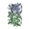
| ||||||||
|---|---|---|---|---|---|---|---|---|---|
| 1 |
| ||||||||
| Unit cell |
|
-Components
| #1: Protein | Mass: 43705.727 Da / Num. of mol.: 2 / Mutation: YES Source method: isolated from a genetically manipulated source Details: CIS PEPTIDE BOND BETWEEN GLYCINE 130 AND SERINE 131 Source: (gene. exp.) BRADYRHIZOBIUM JAPONICUM (bacteria) / Plasmid: PUC18 / Production host: #2: Water | ChemComp-HOH / |  Mass: 18.015 Da / Num. of mol.: 925 / Source method: isolated from a natural source / Formula: H2O Mass: 18.015 Da / Num. of mol.: 925 / Source method: isolated from a natural source / Formula: H2OCompound details | ENGINEERED | |
|---|
-Experimental details
-Experiment
| Experiment | Method: X-RAY DIFFRACTION / Number of used crystals: 1 |
|---|
- Sample preparation
| Crystal | Density Matthews: 2 Å3/Da / Density % sol: 39.9 % |
|---|---|
| Crystal grow | pH: 7 / Details: 20% POLYETHYLENE GLYCOL, 0.1 M TRIS-HCL, PH 7.0 |
-Data collection
| Diffraction | Mean temperature: 100 K |
|---|---|
| Diffraction source | Source: ROTATING ANODE / Type: RIGAKU RU300 / Wavelength: 1.5418 |
| Detector | Type: RIGAKU IMAGE PLATE / Detector: IMAGE PLATE / Details: MIRRORS |
| Radiation | Monochromator: GRAPHITE / Protocol: SINGLE WAVELENGTH / Monochromatic (M) / Laue (L): M / Scattering type: x-ray |
| Radiation wavelength | Wavelength: 1.5418 Å / Relative weight: 1 |
| Reflection | Resolution: 1.9→20 Å / Num. obs: 58083 / % possible obs: 91.7 % / Observed criterion σ(I): 1 / Redundancy: 4.5 % / Rsym value: 0.078 / Net I/σ(I): 13 |
| Reflection shell | Resolution: 1.9→1.97 Å / Redundancy: 2.5 % / Mean I/σ(I) obs: 2.68 / Rsym value: 0.281 / % possible all: 81.3 |
- Processing
| Software |
| ||||||||||||||||||||||||||||||||||||||||||||||||||||||||||||
|---|---|---|---|---|---|---|---|---|---|---|---|---|---|---|---|---|---|---|---|---|---|---|---|---|---|---|---|---|---|---|---|---|---|---|---|---|---|---|---|---|---|---|---|---|---|---|---|---|---|---|---|---|---|---|---|---|---|---|---|---|---|
| Refinement | Method to determine structure: DIRECT METHODS Starting model: PDB ENTRY 1GR8 1gr8 Resolution: 1.9→20 Å / Cross valid method: THROUGHOUT / σ(F): 1 Details: CIS PEPTIDE BOND BETWEEN GLYCINE 130 AND SERINE 131
| ||||||||||||||||||||||||||||||||||||||||||||||||||||||||||||
| Refinement step | Cycle: LAST / Resolution: 1.9→20 Å
| ||||||||||||||||||||||||||||||||||||||||||||||||||||||||||||
| Refine LS restraints |
| ||||||||||||||||||||||||||||||||||||||||||||||||||||||||||||
| Xplor file |
|
