 Movie
Movie Controller

[English] 日本語
 Yorodumi
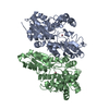
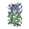
- PDB-4bia: Crystal structure of SCP2 thiolase from Trypanosoma brucei: The C... -
+ Open data

- Basic information
| Entry | Database: PDB / ID: 4bia | ||||||
|---|---|---|---|---|---|---|---|
| Title | Crystal structure of SCP2 thiolase from Trypanosoma brucei: The C337A mutant. | ||||||
 Components Components | 3-KETOACYL-COA THIOLASE, PUTATIVE | ||||||
 Keywords Keywords | TRANSFERASE | ||||||
| Function / homology |  Function and homology information Function and homology informationacetyl-CoA C-acyltransferase / acetyl-CoA C-acyltransferase activity / kinetoplast / acyltransferase activity / lipid transport / lipid binding / mitochondrion Similarity search - Function | ||||||
| Biological species |  | ||||||
| Method |  X-RAY DIFFRACTION / MOLECULAR REPLACEMENT / Resolution: 2.9 Å X-RAY DIFFRACTION / MOLECULAR REPLACEMENT / Resolution: 2.9 Å | ||||||
 Authors Authors | Harijan, R.K. / Kiema, T.-R. / Weiss, M.S. / Michels, P.A.M. / Wierenga, R.K. | ||||||
 Citation Citation | Journal: Biochem.J. / Year: 2013 Title: Crystal Structures of Scp2-Thiolases of Trypanosomatidae, Human Pathogens Causing Widespread Tropical Diseases: The Importance for Catalysis of the Cysteine of the Unique Hdcf Loop. Authors: Harijan, R.K. / Kiema, T.-R. / Karjalainen, M.P. / Janardan, N. / Murthy, M.R.N. / Weiss, M.S. / Michels, P.A.M. / Wierenga, R.K. | ||||||
| History |
|
- Structure visualization
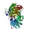
| Structure viewer | Molecule: MolmilJmol/JSmol |
|---|
- Downloads & links
-Download
| PDBx/mmCIF format | 4bia.cif.gz | 312.3 KB | Display | PDBx/mmCIF format |
|---|---|---|---|---|
| PDB format | pdb4bia.ent.gz | 254.1 KB | Display | PDB format |
| PDBx/mmJSON format | 4bia.json.gz | Tree view | PDBx/mmJSON format | |
| Others |  Other downloads Other downloads |
-Validation report
| Arichive directory | https://data.pdbj.org/pub/pdb/validation_reports/bi/4biaftp://data.pdbj.org/pub/pdb/validation_reports/bi/4bia | HTTPS FTP |
|---|
-Related structure data
| Related structure data | 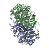 3zbgSC 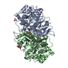 3zbkC 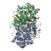 3zblC 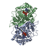 3zbnC 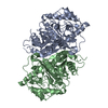 4bi9C S: Starting model for refinement C: citing same article ( |
|---|---|
| Similar structure data |


-Links
 PDBj
PDBj

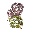
- Assembly
| Deposited unit | 
| ||||||||||||||||||||||||||||||||||||||||||||||||||||||||||
|---|---|---|---|---|---|---|---|---|---|---|---|---|---|---|---|---|---|---|---|---|---|---|---|---|---|---|---|---|---|---|---|---|---|---|---|---|---|---|---|---|---|---|---|---|---|---|---|---|---|---|---|---|---|---|---|---|---|---|---|
| 1 | 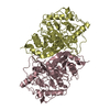
| ||||||||||||||||||||||||||||||||||||||||||||||||||||||||||
| 2 | 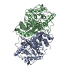
| ||||||||||||||||||||||||||||||||||||||||||||||||||||||||||
| Unit cell |
| ||||||||||||||||||||||||||||||||||||||||||||||||||||||||||
| Noncrystallographic symmetry (NCS) | NCS domain:
NCS domain segments:
NCS oper:
|
-Components
| #1: Protein | Mass: 48073.461 Da / Num. of mol.: 4 / Mutation: YES Source method: isolated from a genetically manipulated source Source: (gene. exp.)  #2: Water | ChemComp-HOH / |  Mass: 18.015 Da / Num. of mol.: 28 / Source method: isolated from a natural source / Formula: H2O Mass: 18.015 Da / Num. of mol.: 28 / Source method: isolated from a natural source / Formula: H2O |
|---|
-Experimental details
-Experiment
| Experiment | Method: X-RAY DIFFRACTION / Number of used crystals: 1 |
|---|
- Sample preparation
| Crystal | Density Matthews: 2.1 Å3/Da / Density % sol: 41.9 % / Description: NONE |
|---|---|
| Crystal grow | pH: 5.5 Details: 100MM BIS TRIS PH5.5, 200MM LITHIUM SULFATE AND 25% PEG3350 |
-Data collection
| Diffraction | Mean temperature: 100 K |
|---|---|
| Diffraction source | Source: ROTATING ANODE / Wavelength: 1.5418 |
| Detector | Type: Bruker Platinum 135 / Detector: CCD / Date: Oct 2, 2012 / Details: HELIOS MIRRORS |
| Radiation | Monochromator: MIRRORS / Protocol: SINGLE WAVELENGTH / Monochromatic (M) / Laue (L): M / Scattering type: x-ray |
| Radiation wavelength | Wavelength: 1.5418 Å / Relative weight: 1 |
| Reflection twin | Operator: -h,-k,l / Fraction: 0.5 |
| Reflection | Resolution: 2.9→41.72 Å / Num. obs: 33721 / % possible obs: 99.9 % / Observed criterion σ(I): 0 / Redundancy: 6.5 % / Rmerge(I) obs: 0.19 / Net I/σ(I): 9.9 |
| Reflection shell | Resolution: 2.9→3 Å / Redundancy: 3.5 % / Rmerge(I) obs: 0.45 / Mean I/σ(I) obs: 1.7 / % possible all: 100 |
- Processing
| Software |
| |||||||||||||||||||||||||||||||||||||||||||||||||||||||||||||||||||||||||||||||||||||||||||
|---|---|---|---|---|---|---|---|---|---|---|---|---|---|---|---|---|---|---|---|---|---|---|---|---|---|---|---|---|---|---|---|---|---|---|---|---|---|---|---|---|---|---|---|---|---|---|---|---|---|---|---|---|---|---|---|---|---|---|---|---|---|---|---|---|---|---|---|---|---|---|---|---|---|---|---|---|---|---|---|---|---|---|---|---|---|---|---|---|---|---|---|---|
| Refinement | Method to determine structure: MOLECULAR REPLACEMENT Starting model: PDB ENTRY 3ZBG Resolution: 2.9→37.363 Å / σ(F): 1.96 / Phase error: 32.73 / Stereochemistry target values: TWIN_LSQ_F Details: THE RESIDUES FROM 1-11, 34-43 AND 102- 104 ARE DISORDERED.
| |||||||||||||||||||||||||||||||||||||||||||||||||||||||||||||||||||||||||||||||||||||||||||
| Solvent computation | Shrinkage radii: 0.9 Å / VDW probe radii: 1.11 Å / Solvent model: FLAT BULK SOLVENT MODEL | |||||||||||||||||||||||||||||||||||||||||||||||||||||||||||||||||||||||||||||||||||||||||||
| Refinement step | Cycle: LAST / Resolution: 2.9→37.363 Å
| |||||||||||||||||||||||||||||||||||||||||||||||||||||||||||||||||||||||||||||||||||||||||||
| Refine LS restraints |
| |||||||||||||||||||||||||||||||||||||||||||||||||||||||||||||||||||||||||||||||||||||||||||
| Refine LS restraints NCS |
| |||||||||||||||||||||||||||||||||||||||||||||||||||||||||||||||||||||||||||||||||||||||||||
| LS refinement shell |
|
