 Movie
Movie Controller


+ Open data

- Basic information
| Entry | Database: PDB / ID: 5tfl | ||||||
|---|---|---|---|---|---|---|---|
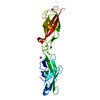
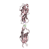
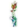
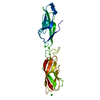
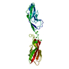
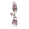
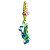
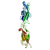
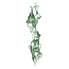
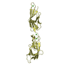
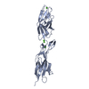
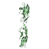
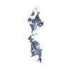
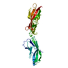
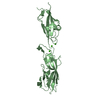
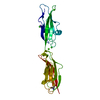
| Title | Crystal Structure of Mouse Cadherin-23 EC7+8 | ||||||
 Components Components | Cadherin-23 | ||||||
 Keywords Keywords | CELL ADHESION / hearing / mechanotransduction / adhesion / calcium-binding protein | ||||||
| Function / homology |  Function and homology information Function and homology informationcochlear hair cell ribbon synapse / equilibrioception / sensory perception of light stimulus / inner ear receptor cell stereocilium organization / stereocilium tip / photoreceptor ribbon synapse / inner ear auditory receptor cell differentiation / kinocilium / calcium-dependent cell-cell adhesion / photoreceptor cell maintenance ...cochlear hair cell ribbon synapse / equilibrioception / sensory perception of light stimulus / inner ear receptor cell stereocilium organization / stereocilium tip / photoreceptor ribbon synapse / inner ear auditory receptor cell differentiation / kinocilium / calcium-dependent cell-cell adhesion / photoreceptor cell maintenance / catenin complex / stereocilium / auditory receptor cell stereocilium organization / inner ear morphogenesis / inner ear development / homophilic cell-cell adhesion / cochlea development / regulation of cytosolic calcium ion concentration / photoreceptor inner segment / sensory perception of sound / locomotory behavior / beta-catenin binding / neuron projection development / apical part of cell / calcium ion transport / cell migration / cell adhesion / cadherin binding / calcium ion binding / centrosome / synapse / plasma membrane Similarity search - Function | ||||||
| Biological species |  | ||||||
| Method |  X-RAY DIFFRACTION / SYNCHROTRON / MOLECULAR REPLACEMENT / Resolution: 3.56 Å X-RAY DIFFRACTION / SYNCHROTRON / MOLECULAR REPLACEMENT / Resolution: 3.56 Å | ||||||
 Authors Authors | Jaiganesh, A. / Sotomayor, M. | ||||||
| Funding support |  United States, 1items United States, 1items
| ||||||
 Citation Citation | Journal: Structure / Year: 2018 Title: Zooming in on Cadherin-23: Structural Diversity and Potential Mechanisms of Inherited Deafness. Authors: Jaiganesh, A. / De-la-Torre, P. / Patel, A.A. / Termine, D.J. / Velez-Cortes, F. / Chen, C. / Sotomayor, M. | ||||||
| History |
|
- Structure visualization
| Structure viewer | Molecule: MolmilJmol/JSmol |
|---|
- Downloads & links
-Download
| PDBx/mmCIF format | 5tfl.cif.gz | 172.7 KB | Display | PDBx/mmCIF format |
|---|---|---|---|---|
| PDB format | pdb5tfl.ent.gz | 137 KB | Display | PDB format |
| PDBx/mmJSON format | 5tfl.json.gz | Tree view | PDBx/mmJSON format | |
| Others |  Other downloads Other downloads |
-Validation report
| Arichive directory | https://data.pdbj.org/pub/pdb/validation_reports/tf/5tflftp://data.pdbj.org/pub/pdb/validation_reports/tf/5tfl | HTTPS FTP |
|---|
-Related structure data
| Related structure data | 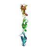 5i8dSC  5tfkC 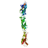 5tfmC  5uluC  5un2C 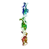 5uz8C  5vh2C 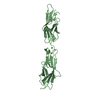 5vt8C  5vvmC 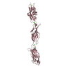 5w4tC  5wj8C  5wjmC C: citing same article ( S: Starting model for refinement |
|---|---|
| Similar structure data |


-Links
 PDBj
PDBj
- Assembly
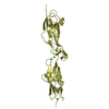
| Deposited unit | 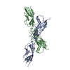
| ||||||||||||||||||
|---|---|---|---|---|---|---|---|---|---|---|---|---|---|---|---|---|---|---|---|
| 1 | 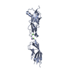
| ||||||||||||||||||
| 2 | 
| ||||||||||||||||||
| Unit cell |
| ||||||||||||||||||
| Noncrystallographic symmetry (NCS) | NCS domain:
NCS domain segments: Component-ID: _ / Ens-ID: 1 / Beg auth comp-ID: PRO / Beg label comp-ID: PRO / End auth comp-ID: LEU / End label comp-ID: LEU / Refine code: _ / Auth seq-ID: 645 - 860 / Label seq-ID: 4 - 219
|
-Components
| #1: Protein | Mass: 25528.568 Da / Num. of mol.: 2 / Fragment: Cadherin domains 7-8, residues 666-886 Source method: isolated from a genetically manipulated source Source: (gene. exp.)  #2: Chemical |   Mass: 22.990 Da / Num. of mol.: 2 / Source method: obtained synthetically / Formula: Na Mass: 22.990 Da / Num. of mol.: 2 / Source method: obtained synthetically / Formula: Na#3: Chemical | ChemComp-CA /   Mass: 40.078 Da / Num. of mol.: 4 / Source method: obtained synthetically / Formula: Ca Mass: 40.078 Da / Num. of mol.: 4 / Source method: obtained synthetically / Formula: Ca#4: Water | ChemComp-HOH / |  Mass: 18.015 Da / Num. of mol.: 3 / Source method: isolated from a natural source / Formula: H2O Mass: 18.015 Da / Num. of mol.: 3 / Source method: isolated from a natural source / Formula: H2OHas protein modification | Y | |
|---|
-Experimental details
-Experiment
| Experiment | Method: X-RAY DIFFRACTION / Number of used crystals: 1 |
|---|
- Sample preparation
| Crystal | Density Matthews: 5.24 Å3/Da / Density % sol: 76.53 % |
|---|---|
| Crystal grow | Temperature: 277 K / Method: vapor diffusion, sitting drop / pH: 4.7 Details: 0.1 M Sodium Acetate pH 4.7, 1.9 M Sodium Chloride, 30% PEG 400 |
-Data collection
| Diffraction | Mean temperature: 100 K / Ambient temp details: Oxford Cryo-Jet crystal cryocoolers |
|---|---|
| Diffraction source | Source: SYNCHROTRON / Site: APS / Beamline: 24-ID-C / Wavelength: 0.97921 Å |
| Detector | Type: DECTRIS PILATUS 6M-F / Detector: PIXEL / Date: Mar 22, 2015 |
| Radiation | Protocol: SINGLE WAVELENGTH / Monochromatic (M) / Laue (L): M / Scattering type: x-ray |
| Radiation wavelength | Wavelength: 0.97921 Å / Relative weight: 1 |
| Reflection | Resolution: 3.56→131.39 Å / Num. obs: 13716 / % possible obs: 100 % / Redundancy: 35.5 % / Biso Wilson estimate: 87.2 Å2 / Rmerge(I) obs: 0.363 / Net I/σ(I): 20 |
| Reflection shell | Resolution: 3.56→3.61 Å / Redundancy: 23.7 % / Rmerge(I) obs: 1.587 / Mean I/σ(I) obs: 2.5 / CC1/2: 0.748 / % possible all: 100 |
- Processing
| Software |
| ||||||||||||||||||||||||||||||||||||||||||||||||||||||||||||||||||||||||||||||||||||||||||||||||||||||||||||||||||||||||||||||||||||||||||||||||||||||||||||||||||||||||||||||||||||||
|---|---|---|---|---|---|---|---|---|---|---|---|---|---|---|---|---|---|---|---|---|---|---|---|---|---|---|---|---|---|---|---|---|---|---|---|---|---|---|---|---|---|---|---|---|---|---|---|---|---|---|---|---|---|---|---|---|---|---|---|---|---|---|---|---|---|---|---|---|---|---|---|---|---|---|---|---|---|---|---|---|---|---|---|---|---|---|---|---|---|---|---|---|---|---|---|---|---|---|---|---|---|---|---|---|---|---|---|---|---|---|---|---|---|---|---|---|---|---|---|---|---|---|---|---|---|---|---|---|---|---|---|---|---|---|---|---|---|---|---|---|---|---|---|---|---|---|---|---|---|---|---|---|---|---|---|---|---|---|---|---|---|---|---|---|---|---|---|---|---|---|---|---|---|---|---|---|---|---|---|---|---|---|---|
| Refinement | Method to determine structure: MOLECULAR REPLACEMENT Starting model: Cadherin-23 (5I8D) Resolution: 3.56→131.39 Å / Cor.coef. Fo:Fc: 0.928 / Cor.coef. Fo:Fc free: 0.921 / SU B: 41.635 / SU ML: 0.266 / Cross valid method: THROUGHOUT / ESU R: 1.781 / ESU R Free: 0.395 / Details: HYDROGENS HAVE BEEN ADDED IN THE RIDING POSITIONS
| ||||||||||||||||||||||||||||||||||||||||||||||||||||||||||||||||||||||||||||||||||||||||||||||||||||||||||||||||||||||||||||||||||||||||||||||||||||||||||||||||||||||||||||||||||||||
| Solvent computation | Ion probe radii: 0.8 Å / Shrinkage radii: 0.8 Å / VDW probe radii: 1.2 Å | ||||||||||||||||||||||||||||||||||||||||||||||||||||||||||||||||||||||||||||||||||||||||||||||||||||||||||||||||||||||||||||||||||||||||||||||||||||||||||||||||||||||||||||||||||||||
| Displacement parameters | Biso mean: 90.48 Å2
| ||||||||||||||||||||||||||||||||||||||||||||||||||||||||||||||||||||||||||||||||||||||||||||||||||||||||||||||||||||||||||||||||||||||||||||||||||||||||||||||||||||||||||||||||||||||
| Refinement step | Cycle: 1 / Resolution: 3.56→131.39 Å
| ||||||||||||||||||||||||||||||||||||||||||||||||||||||||||||||||||||||||||||||||||||||||||||||||||||||||||||||||||||||||||||||||||||||||||||||||||||||||||||||||||||||||||||||||||||||
| Refine LS restraints |
|