 Movie
Movie Controller


+ Open data

- Basic information
| Entry | Database: PDB / ID: 4zmz | ||||||
|---|---|---|---|---|---|---|---|
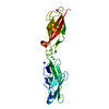
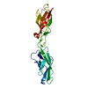
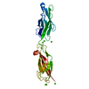
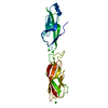
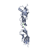
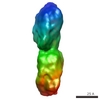
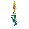
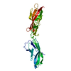
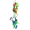
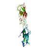
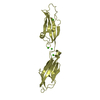
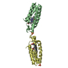
| Title | Crystal structure of human P-cadherin (monomer 2) | ||||||
 Components Components | Cadherin-3 | ||||||
 Keywords Keywords | CELL ADHESION / dimerization / conformational change | ||||||
| Function / homology |  Function and homology information Function and homology informationnegative regulation of timing of catagen / hair cycle process / regulation of transport / positive regulation of keratinocyte proliferation / positive regulation of melanin biosynthetic process / calcium-dependent cell-cell adhesion / hair follicle maturation / cell-cell adhesion mediated by cadherin / adherens junction organization / catenin complex ...negative regulation of timing of catagen / hair cycle process / regulation of transport / positive regulation of keratinocyte proliferation / positive regulation of melanin biosynthetic process / calcium-dependent cell-cell adhesion / hair follicle maturation / cell-cell adhesion mediated by cadherin / adherens junction organization / catenin complex / retina homeostasis / cell-cell junction assembly / positive regulation of insulin-like growth factor receptor signaling pathway / Adherens junctions interactions / keratinization / homophilic cell-cell adhesion / visual perception / adherens junction / negative regulation of transforming growth factor beta receptor signaling pathway / beta-catenin binding / cell morphogenesis / cell junction / positive regulation of canonical Wnt signaling pathway / cell migration / cell adhesion / cadherin binding / calcium ion binding / plasma membrane / cytoplasm Similarity search - Function | ||||||
| Biological species |  Homo sapiens (human) Homo sapiens (human) | ||||||
| Method |  X-RAY DIFFRACTION / SYNCHROTRON / MOLECULAR REPLACEMENT / Resolution: 2.05 Å X-RAY DIFFRACTION / SYNCHROTRON / MOLECULAR REPLACEMENT / Resolution: 2.05 Å | ||||||
 Authors Authors | Caaveiro, J.M.M. / Kudo, S. / Tsumoto, K. | ||||||
 Citation Citation | Journal: Structure / Year: 2016 Title: Adhesive Dimerization of Human P-Cadherin Catalyzed by a Chaperone-like Mechanism Authors: Kudo, S. / Caaveiro, J.M. / Tsumoto, K. | ||||||
| History |
|
- Structure visualization
| Structure viewer | Molecule: MolmilJmol/JSmol |
|---|
- Downloads & links
-Download
| PDBx/mmCIF format | 4zmz.cif.gz | 59.6 KB | Display | PDBx/mmCIF format |
|---|---|---|---|---|
| PDB format | pdb4zmz.ent.gz | 41.1 KB | Display | PDB format |
| PDBx/mmJSON format | 4zmz.json.gz | Tree view | PDBx/mmJSON format | |
| Others |  Other downloads Other downloads |
-Validation report
| Arichive directory | https://data.pdbj.org/pub/pdb/validation_reports/zm/4zmzftp://data.pdbj.org/pub/pdb/validation_reports/zm/4zmz | HTTPS FTP |
|---|
-Related structure data
| Related structure data |  4zmlC  4zmnSC  4zmoC  4zmpC  4zmqC  4zmtC  4zmvC  4zmwC  4zmxC  4zmyC C: citing same article ( S: Starting model for refinement |
|---|---|
| Similar structure data |

-Links
 PDBj
PDBj

- Assembly
| Deposited unit | 
| ||||||||
|---|---|---|---|---|---|---|---|---|---|
| 1 |
| ||||||||
| Unit cell |
|
-Components
| #1: Protein | Mass: 23448.008 Da / Num. of mol.: 1 / Fragment: UNP residues 107-320 Source method: isolated from a genetically manipulated source Source: (gene. exp.) Homo sapiens (human) / Gene: CDH3, CDHP / Plasmid: Champion pET SUMO / Production host:  | ||
|---|---|---|---|
| #2: Chemical | ChemComp-CA /   Mass: 40.078 Da / Num. of mol.: 4 / Source method: obtained synthetically / Formula: Ca Mass: 40.078 Da / Num. of mol.: 4 / Source method: obtained synthetically / Formula: Ca#3: Water | ChemComp-HOH / |  Mass: 18.015 Da / Num. of mol.: 91 / Source method: isolated from a natural source / Formula: H2O Mass: 18.015 Da / Num. of mol.: 91 / Source method: isolated from a natural source / Formula: H2O |
-Experimental details
-Experiment
| Experiment | Method: X-RAY DIFFRACTION |
|---|
- Sample preparation
| Crystal | Density Matthews: 2.39 Å3/Da / Density % sol: 48.46 % |
|---|---|
| Crystal grow | Temperature: 293 K / Method: vapor diffusion, hanging drop / pH: 7.5 / Details: 28% PEG 400 200 mM CaCl2 95 mM HEPES |
-Data collection
| Diffraction | Mean temperature: 100 K |
|---|---|
| Diffraction source | Source: SYNCHROTRON / Site: Photon Factory  / Beamline: AR-NW12A / Wavelength: 1 Å / Beamline: AR-NW12A / Wavelength: 1 Å |
| Detector | Type: ADSC QUANTUM 210r / Detector: CCD / Date: Jun 24, 2013 |
| Radiation | Protocol: SINGLE WAVELENGTH / Monochromatic (M) / Laue (L): M / Scattering type: x-ray |
| Radiation wavelength | Wavelength: 1 Å / Relative weight: 1 |
| Reflection | Resolution: 2.05→37.7 Å / Num. obs: 13000 / % possible obs: 92.2 % / Redundancy: 3.3 % / Rmerge(I) obs: 0.081 / Net I/σ(I): 9.4 |
| Reflection shell | Resolution: 2.05→2.16 Å / Redundancy: 3.3 % / Rmerge(I) obs: 0.288 / Mean I/σ(I) obs: 3.3 / % possible all: 83.4 |
- Processing
| Software |
| ||||||||||||||||||||||||||||||||||||||||||||||||||||||||||||||||||||||||||||||||||||||||||||||||||||||||||||||||||||||||||||||||||||||||||||||||||||||||||||||||||||||||||||||||||||||
|---|---|---|---|---|---|---|---|---|---|---|---|---|---|---|---|---|---|---|---|---|---|---|---|---|---|---|---|---|---|---|---|---|---|---|---|---|---|---|---|---|---|---|---|---|---|---|---|---|---|---|---|---|---|---|---|---|---|---|---|---|---|---|---|---|---|---|---|---|---|---|---|---|---|---|---|---|---|---|---|---|---|---|---|---|---|---|---|---|---|---|---|---|---|---|---|---|---|---|---|---|---|---|---|---|---|---|---|---|---|---|---|---|---|---|---|---|---|---|---|---|---|---|---|---|---|---|---|---|---|---|---|---|---|---|---|---|---|---|---|---|---|---|---|---|---|---|---|---|---|---|---|---|---|---|---|---|---|---|---|---|---|---|---|---|---|---|---|---|---|---|---|---|---|---|---|---|---|---|---|---|---|---|---|
| Refinement | Method to determine structure: MOLECULAR REPLACEMENT Starting model: 4ZMN Resolution: 2.05→32.42 Å / Cor.coef. Fo:Fc: 0.941 / Cor.coef. Fo:Fc free: 0.917 / SU B: 7.915 / SU ML: 0.199 / Cross valid method: THROUGHOUT / ESU R: 0.265 / ESU R Free: 0.215 / Stereochemistry target values: MAXIMUM LIKELIHOOD / Details: HYDROGENS HAVE BEEN ADDED IN THE RIDING POSITIONS
| ||||||||||||||||||||||||||||||||||||||||||||||||||||||||||||||||||||||||||||||||||||||||||||||||||||||||||||||||||||||||||||||||||||||||||||||||||||||||||||||||||||||||||||||||||||||
| Solvent computation | Ion probe radii: 0.8 Å / Shrinkage radii: 0.8 Å / VDW probe radii: 1.2 Å / Solvent model: BABINET MODEL WITH MASK | ||||||||||||||||||||||||||||||||||||||||||||||||||||||||||||||||||||||||||||||||||||||||||||||||||||||||||||||||||||||||||||||||||||||||||||||||||||||||||||||||||||||||||||||||||||||
| Displacement parameters | Biso mean: 32.861 Å2
| ||||||||||||||||||||||||||||||||||||||||||||||||||||||||||||||||||||||||||||||||||||||||||||||||||||||||||||||||||||||||||||||||||||||||||||||||||||||||||||||||||||||||||||||||||||||
| Refinement step | Cycle: 1 / Resolution: 2.05→32.42 Å
| ||||||||||||||||||||||||||||||||||||||||||||||||||||||||||||||||||||||||||||||||||||||||||||||||||||||||||||||||||||||||||||||||||||||||||||||||||||||||||||||||||||||||||||||||||||||
| Refine LS restraints |
|