 Movie
Movie Controller

[English] 日本語
 Yorodumi
- PDB-5t48: Crystal structure of the D. melanogaster eIF4E-eIF4G complex with... -
+ Open data

- Basic information
| Entry | Database: PDB / ID: 5t48 | ||||||
|---|---|---|---|---|---|---|---|
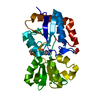
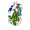
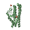
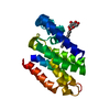
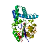
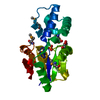
| Title | Crystal structure of the D. melanogaster eIF4E-eIF4G complex without lateral contact | ||||||
 Components Components |
| ||||||
 Keywords Keywords | TRANSLATION / GENE REGULATION / CAP BINDING PROTEIN / 4E-BINDING PROTEIN / TRANSLATION INITIATION / eIF4F | ||||||
| Function / homology |  Function and homology information Function and homology informationTOR signaling pathway / : / Activation of the mRNA upon binding of the cap-binding complex and eIFs, and subsequent binding to 43S / Transport of the SLBP independent Mature mRNA / Transport of the SLBP Dependant Mature mRNA / Transport of Mature mRNA Derived from an Intronless Transcript / L13a-mediated translational silencing of Ceruloplasmin expression / mTORC1-mediated signalling / Translation initiation complex formation / Ribosomal scanning and start codon recognition ...TOR signaling pathway / : / Activation of the mRNA upon binding of the cap-binding complex and eIFs, and subsequent binding to 43S / Transport of the SLBP independent Mature mRNA / Transport of the SLBP Dependant Mature mRNA / Transport of Mature mRNA Derived from an Intronless Transcript / L13a-mediated translational silencing of Ceruloplasmin expression / mTORC1-mediated signalling / Translation initiation complex formation / Ribosomal scanning and start codon recognition / muscle cell postsynaptic specialization / RNA metabolic process / neuronal ribonucleoprotein granule / eukaryotic initiation factor 4G binding / eukaryotic initiation factor 4E binding / RNA cap binding / eukaryotic translation initiation factor 4F complex / RNA 7-methylguanosine cap binding / translation initiation factor activity / neuromuscular junction / translational initiation / P-body / mitotic cell cycle / regulation of translation / nuclear body / translation / RNA binding / cytosol / cytoplasm Similarity search - Function | ||||||
| Biological species |  | ||||||
| Method |  X-RAY DIFFRACTION / SYNCHROTRON / MOLECULAR REPLACEMENT / Resolution: 2.19 Å X-RAY DIFFRACTION / SYNCHROTRON / MOLECULAR REPLACEMENT / Resolution: 2.19 Å | ||||||
 Authors Authors | Gruener, S. / Peter, D. / Weber, R. / Wohlbold, L. / Chung, M.-Y. / Weichenrieder, O. / Valkov, E. / Igreja, C. / Izaurralde, E. | ||||||
 Citation Citation | Journal: Mol.Cell / Year: 2016 Title: The Structures of eIF4E-eIF4G Complexes Reveal an Extended Interface to Regulate Translation Initiation. Authors: Gruner, S. / Peter, D. / Weber, R. / Wohlbold, L. / Chung, M.Y. / Weichenrieder, O. / Valkov, E. / Igreja, C. / Izaurralde, E. | ||||||
| History |
| ||||||
| Remark 650 | HELIX DETERMINATION METHOD: AUTHOR PROVIDED. | ||||||
| Remark 700 | SHEET DETERMINATION METHOD: AUTHOR PROVIDED. |
- Structure visualization
| Structure viewer | Molecule: MolmilJmol/JSmol |
|---|
- Downloads & links
-Download
| PDBx/mmCIF format | 5t48.cif.gz | 147.1 KB | Display | PDBx/mmCIF format |
|---|---|---|---|---|
| PDB format | pdb5t48.ent.gz | 117.4 KB | Display | PDB format |
| PDBx/mmJSON format | 5t48.json.gz | Tree view | PDBx/mmJSON format | |
| Others |  Other downloads Other downloads |
-Validation report
| Arichive directory | https://data.pdbj.org/pub/pdb/validation_reports/t4/5t48ftp://data.pdbj.org/pub/pdb/validation_reports/t4/5t48 | HTTPS FTP |
|---|
-Related structure data
| Related structure data | 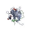 5t46C  5t47C 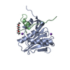 4ue8S S: Starting model for refinement C: citing same article ( |
|---|---|
| Similar structure data |


-Links
 PDBj
PDBj

- Assembly
| Deposited unit | 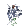
| ||||||||
|---|---|---|---|---|---|---|---|---|---|
| 1 |
| ||||||||
| Unit cell |
|
-Components
| #1: Protein | Mass: 21249.037 Da / Num. of mol.: 1 Source method: isolated from a genetically manipulated source Source: (gene. exp.)  |
|---|---|
| #2: Protein | Mass: 7410.389 Da / Num. of mol.: 1 Source method: isolated from a genetically manipulated source Source: (gene. exp.) |
| #3: Chemical | ChemComp-MGP /   Mass: 538.215 Da / Num. of mol.: 1 / Source method: obtained synthetically / Formula: C11H19N5O14P3 Mass: 538.215 Da / Num. of mol.: 1 / Source method: obtained synthetically / Formula: C11H19N5O14P3 |
| #4: Water | ChemComp-HOH /  Mass: 18.015 Da / Num. of mol.: 35 / Source method: isolated from a natural source / Formula: H2O Mass: 18.015 Da / Num. of mol.: 35 / Source method: isolated from a natural source / Formula: H2O |
-Experimental details
-Experiment
| Experiment | Method: X-RAY DIFFRACTION / Number of used crystals: 1 |
|---|
- Sample preparation
| Crystal | Density Matthews: 2 Å3/Da / Density % sol: 39 % |
|---|---|
| Crystal grow | Temperature: 291 K / Method: vapor diffusion, hanging drop / pH: 8 Details: 0.1 M TrisHCl pH 8.0, 0.01 M ZnCl2, 20% w/v PEG6000 |
-Data collection
| Diffraction | Mean temperature: 100 K |
|---|---|
| Diffraction source | Source: SYNCHROTRON / Site: SLS  / Beamline: X10SA / Wavelength: 0.99992 Å / Beamline: X10SA / Wavelength: 0.99992 Å |
| Detector | Type: DECTRIS PILATUS 6M / Detector: PIXEL / Date: Apr 19, 2015 / Details: DYNAMICALLY BENDABLE MIRRORS |
| Radiation | Monochromator: Si(111) / Protocol: SINGLE WAVELENGTH / Monochromatic (M) / Laue (L): M / Scattering type: x-ray |
| Radiation wavelength | Wavelength: 0.99992 Å / Relative weight: 1 |
| Reflection | Resolution: 2.19→45.62 Å / Num. obs: 12336 / % possible obs: 100 % / Observed criterion σ(I): -3 / Redundancy: 12.6 % / Biso Wilson estimate: 46.84 Å2 / CC1/2: 0.999 / Rsym value: 0.081 / Net I/σ(I): 17 |
| Reflection shell | Resolution: 2.19→2.26 Å / Redundancy: 11.4 % / Mean I/σ(I) obs: 2.3 / CC1/2: 0.85 / Rsym value: 1.066 / % possible all: 99.6 |
- Processing
| Software |
| ||||||||||||||||||||||||||||||||||||||||||||||||||||||||||||||||||||||||||||||||||||||||||||||||||||||||||||||||||||||||||||||||||||||||||||||||||||||
|---|---|---|---|---|---|---|---|---|---|---|---|---|---|---|---|---|---|---|---|---|---|---|---|---|---|---|---|---|---|---|---|---|---|---|---|---|---|---|---|---|---|---|---|---|---|---|---|---|---|---|---|---|---|---|---|---|---|---|---|---|---|---|---|---|---|---|---|---|---|---|---|---|---|---|---|---|---|---|---|---|---|---|---|---|---|---|---|---|---|---|---|---|---|---|---|---|---|---|---|---|---|---|---|---|---|---|---|---|---|---|---|---|---|---|---|---|---|---|---|---|---|---|---|---|---|---|---|---|---|---|---|---|---|---|---|---|---|---|---|---|---|---|---|---|---|---|---|---|---|---|---|
| Refinement | Method to determine structure: MOLECULAR REPLACEMENT Starting model: 4UE8 Resolution: 2.19→39.063 Å / SU ML: 0.23 / Cross valid method: FREE R-VALUE / σ(F): 1.36 / Phase error: 26.16 / Details: HYDROGENS WERE REFINED IN THE RIDING POSITIONS.
| ||||||||||||||||||||||||||||||||||||||||||||||||||||||||||||||||||||||||||||||||||||||||||||||||||||||||||||||||||||||||||||||||||||||||||||||||||||||
| Solvent computation | Shrinkage radii: 0.9 Å / VDW probe radii: 1.11 Å | ||||||||||||||||||||||||||||||||||||||||||||||||||||||||||||||||||||||||||||||||||||||||||||||||||||||||||||||||||||||||||||||||||||||||||||||||||||||
| Displacement parameters | Biso mean: 70.66 Å2 | ||||||||||||||||||||||||||||||||||||||||||||||||||||||||||||||||||||||||||||||||||||||||||||||||||||||||||||||||||||||||||||||||||||||||||||||||||||||
| Refinement step | Cycle: LAST / Resolution: 2.19→39.063 Å
| ||||||||||||||||||||||||||||||||||||||||||||||||||||||||||||||||||||||||||||||||||||||||||||||||||||||||||||||||||||||||||||||||||||||||||||||||||||||
| Refine LS restraints |
| ||||||||||||||||||||||||||||||||||||||||||||||||||||||||||||||||||||||||||||||||||||||||||||||||||||||||||||||||||||||||||||||||||||||||||||||||||||||
| LS refinement shell |
| ||||||||||||||||||||||||||||||||||||||||||||||||||||||||||||||||||||||||||||||||||||||||||||||||||||||||||||||||||||||||||||||||||||||||||||||||||||||
| Refinement TLS params. | Method: refined / Refine-ID: X-RAY DIFFRACTION
| ||||||||||||||||||||||||||||||||||||||||||||||||||||||||||||||||||||||||||||||||||||||||||||||||||||||||||||||||||||||||||||||||||||||||||||||||||||||
| Refinement TLS group |
|
